A Brief Introduction to Muscles
Muscles produce the forces for movement and stabilization of the human body. When movement occurs against a resisting or restraining force, energy is expended by muscles, and energy must be supplied to the muscles. Understanding muscle structure and function is essential for analyzing laryngeal biomechanics.
Types of Muscle
Because the human body performs so many different motor functions, different types of muscles have developed. Each muscle type evolved to perform specific tasks.
Three Major Types
Striated (Skeletal) Muscle:
- Used for movement and posturing
- Attached to bones via tendons
- Voluntary control
- Focus of this section
Smooth Muscle:
- Used around intestines and blood vessels
- Propagates fluids through organs
- Involuntary control
- Slower contraction
Heart Muscle (Cardiac):
- Used to pump blood
- Specialized for continuous rhythmic contraction
- Involuntary control
- Never fatigues under normal conditions
Muscle Specialization by Function
Each muscle develops and evolves to perform a specific task, with structure matched to function.
Postural Muscles
Function: Hold tissues together, resist gravity, stabilize skeletal framework
Requirements:
- High resistance to fatigue (seldom get rest)
- Precise force control (gentle balancing)
- Moderate force range (not maximum strength)
Example: Neck muscles steadying the head when upright
- Continuous low-level activation
- Fine adjustments for positioning
- Accuracy more important than maximum strength
Movement Muscles
Function: Execute quick movements
Requirements:
- Fast response time
- High power output
- Can afford recovery time between contractions
- Burst energy expenditure
Example: Eye blink
- Extremely rapid contraction
- Brief duration
- Frequent rest periods between blinks
Laryngeal Muscle Demands
Unique challenge: Laryngeal muscles must serve dual functions
- Respiration: Continuous, fatigue-resistant (like postural muscles)
- Phonation: Quick response (like movement muscles)
This dual demand results in specialized fiber composition discussed in the next section.
Hierarchical Structure of Muscle
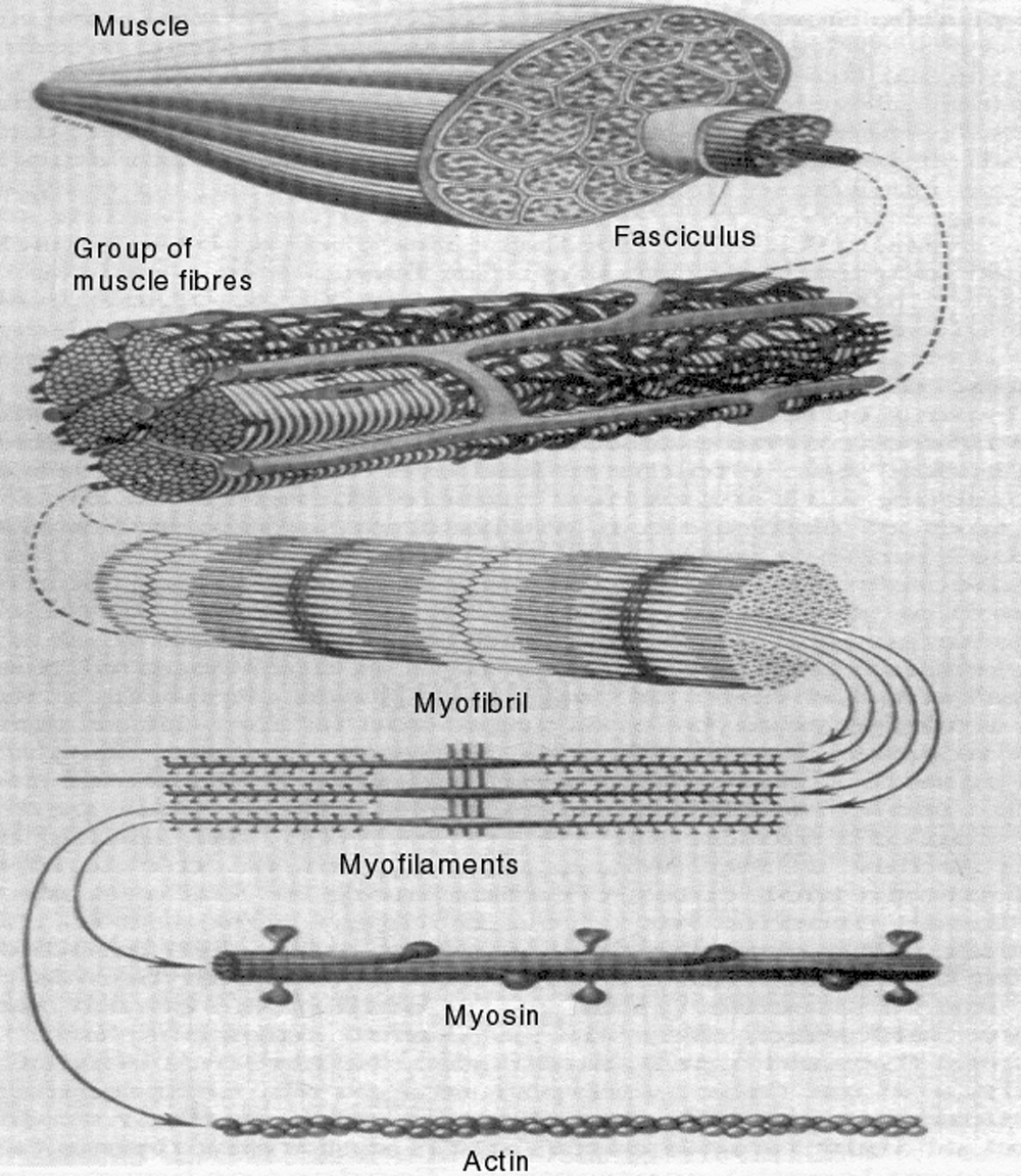 Figure 2.11: Structural components of muscle from whole muscle down to molecular level.
Muscle exhibits hierarchical organization from macroscopic to molecular scales.
Level 1: Muscle Fascicles
Muscle fascicles comprise groups of muscle fibers:
- Enclosed by a connective-tissue sheath called the perimysium (the whole muscle is wrapped in epimysium, and each fiber in endomysium); the sarcoplasmic reticulum is something different—the calcium-storing membrane network inside each fiber
- Each fascicle contains numerous individual fibers
- Blood vessels and nerves surround fiber groups
- Visible to naked eye as bundles within whole muscle
Level 2: Muscle Fibers
Muscle fibers are the basic functional units:
- Barely visible to naked eye (~0.1 mm diameter)
- Length can extend along entire muscle
- Each fiber contains numerous myofibrils
- Enclosed by cell membrane (sarcolemma)
Level 3: Myofibrils
Myofibrils are smaller structural units within each fiber:
- Diameter ~1-2 micrometers
- Run length of fiber
- Contains repeating pattern of sarcomeres
- Site of actual contraction
Level 4: Myofilaments
Myofilaments are the components of myofibrils:
- Two types: thick filaments (myosin) and thin filaments (actin)
- Arranged in overlapping pattern
- Interaction between filaments produces contraction
Level 5: Molecular
At the molecular level:
- Myosin molecules: Form thick filaments
- Actin molecules: Form thin filaments
- Cross-bridges: Projections from myosin that interact with actin
- This is where force is generated
The Sliding Filament Theory
Muscle contraction can be explained by the sliding filament theory, developed in the 1950s.
Basic Mechanism
Within each sarcomere (repeating unit of myofibril):
- Actin filaments slide past myosin filaments
- Creates more or less overlap
- Changes sarcomere length without changing filament lengths
The Cross-Bridge Cycle
Cross bridges (protrusions from myosin) propel the sliding:
Step 1: Attachment
- Cross bridge binds to actin filament
- Forms actin-myosin complex
Step 2: Power Stroke
- Cross bridge bends (like rowing motion)
- Pulls actin filament past myosin
- Uses energy from ATP breakdown
Step 3: Detachment
- Cross bridge releases from actin
- Requires new ATP molecule
Step 4: Recovery
- Cross bridge returns to original position
- Ready to attach again
Analogy: Like oars extending from edge of boat
- Oar reaches forward (recovery)
- Oar grabs water (attachment)
- Oar pulls backward (power stroke)
- Oar releases water (detachment)
- Cycle repeats
Limit of Contraction
Internal contraction is limited by:
- Amount of space (non-overlap) between filament segments
- Maximum shortening: ~50-60% of resting length
- Optimal overlap for maximum force generation
Microscopic Versus Macroscopic Contraction
Isometric Contraction
Isometric means “constant length”:
- Microscopic: Sarcomeres contract, filaments slide
- Macroscopic: Overall muscle length stays constant
- How: Tendons or ligaments at muscle ends stretch
Why this is important:
- Microscopic contraction can occur without apparent macroscopic contraction
- Allows internal force generation while maintaining position
- Used to study maximum forces muscle can produce
Isotonic Contraction
Isotonic means “constant tension”:
- Force remains constant
- Length changes
- Typical of most voluntary movements
Energy Requirements
ATP: The Energy Currency
Muscle contraction requires:
- ATP (adenosine triphosphate)
- Split into ADP + phosphate
- Releases energy for cross-bridge cycle
- Must be continuously resupplied
Metabolic Pathways
Aerobic metabolism (with oxygen):
- Efficient energy production
- Sustainable for long periods
- Used by fatigue-resistant fibers
Anaerobic metabolism (without oxygen):
- Rapid energy production
- Cannot be sustained
- Used by fast-twitch fibers
- Produces lactate (causes fatigue)
Laryngeal Muscle Considerations
Continuous Function
Laryngeal muscles face unique challenge:
- Must function continuously for respiration
- Cannot completely rest
- Requires high fatigue resistance
Rapid Response
Simultaneously, laryngeal muscles need:
- Quick reflex protection of airway
- Rapid adjustments for phonation
- Speed of response
Fiber Type Implications
These demands result in:
- Special mixture of fiber types
- High proportion of fast, fatigue-resistant fibers
- Discussed in detail in next section on muscle fiber typing
Summary
Muscles are hierarchically organized from molecular to macroscopic scales:
Structure:
- Whole muscle → Fascicles → Fibers → Myofibrils → Myofilaments → Molecules
Contraction Mechanism:
- Sliding of actin past myosin filaments
- Powered by cross-bridge cycles
- Requires ATP energy
Types:
- Striated (skeletal): Voluntary movement
- Smooth: Involuntary organ function
- Cardiac: Heart contraction
Muscle Specialization:
- Postural muscles: Fatigue-resistant, continuous low force
- Movement muscles: Fast, powerful, intermittent
- Laryngeal muscles: Both fast and fatigue-resistant
Isometric Versus Isotonic:
- Isometric: Constant length, changing force
- Isotonic: Constant force, changing length
Understanding muscle structure and function provides foundation for:
- Analyzing laryngeal muscle biomechanics
- Understanding force generation
- Explaining fatigue and training effects
- Designing therapeutic interventions
Key Takeaways
- ✅ Three muscle types exist: striated (voluntary movement), smooth (organs), and cardiac (heart)
- ✅ Muscle has hierarchical structure from whole muscle down to molecular filaments
- ✅ Sliding filament theory explains contraction: actin slides past myosin via cross-bridge cycles
- ✅ Isometric contraction (constant length) can occur at microscopic level while macroscopic length stays constant
- ✅ Laryngeal muscles must be both fast-responding and fatigue-resistant due to dual respiratory/phonatory function
- ✅ ATP provides energy for cross-bridge cycle and must be continuously resupplied
Related Topics
Further Reading
- Burke, R. E. (1981). Motor units: Anatomy, physiology, and functional organization. In V. B. Brooks (Ed.), Motor control (Handbook of physiology, Section 1, Vol. 2, Pt. 1), pp. 506-512. Bethesda: American Physiological Society.
- Aidley, D. J. (1989). The physiology of excitable cells (3rd Ed). Cambridge: Cambridge University Press.