Mechanics of Vocal Fold Elongation
Vocal fold length is the primary geometric variable for adjustment of fundamental frequency. Understanding how the laryngeal cartilages move relative to one another, and how muscles produce these movements, is essential for predicting F₀ control. The analogy to stringed instruments provides useful context: in a violin, pegs adjust string tension and finger positions alter length. In the vocal folds, stress and length are inextricably linked—length increase is the primary means by which fibers in tissue layers become stressed.
The Laryngeal Framework Revisited
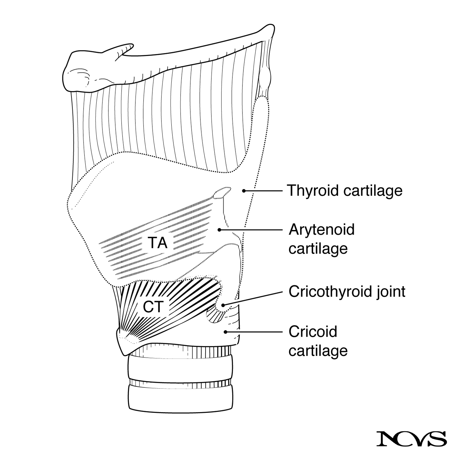 Figure 8.1: Cartilaginous framework of the larynx in lateral aspect, showing cricothyroid (CT) and thyroarytenoid (TA) muscles.
The laryngeal framework comprises several articulated cartilages:
Primary Structures
- Thyroid cartilage: Large shield-like structure forming anterior and lateral walls
- Cricoid cartilage: Complete ring forming foundation of larynx
- Arytenoid cartilages: Pyramidal structures sitting atop posterior cricoid
- Cricothyroid joint: Articulation allowing rotation and some translation
Movement Capabilities
The cricothyroid joint permits two types of movement:
Rotation
- Cricoid rocks backward (or thyroid rocks forward)
- Reduces cricothyroid space anteriorly
- Arytenoid cartilages follow backward movement of cricoid
- Results in vocal fold elongation
Translation
- Forward sliding of thyroid on cricoid (or vice versa)
- Provides additional elongation beyond rotation alone
- Magnitude depends on joint morphology
- Individual variation in translation capability
Simplified Mechanical Model
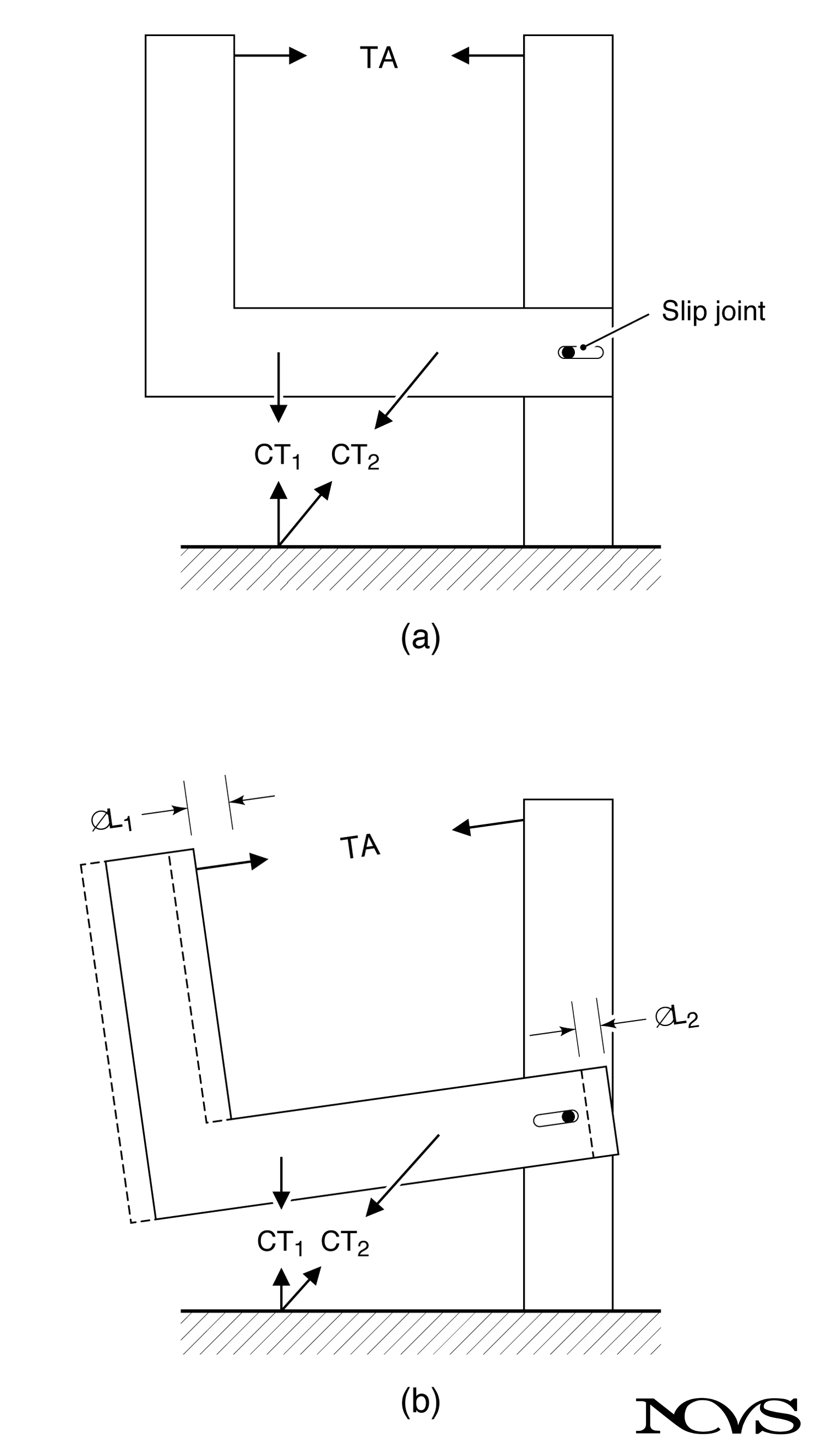 Figure 8.2: Schematic representation of major laryngeal cartilages in rotation about the slipping cricothyroid joint. CT₁, CT₂, and TA show the contractile forces of the cricothyroid (pars recta and pars obliqua) and thyroarytenoid muscles, respectively.
A simplified model clarifies the mechanical principles:
Model Components
- Vertical rod: Combined cricoid and arytenoid cartilages (anchored to ground)
- Movable elbow: Thyroid cartilage
- Slip joint: Cricothyroid articulation allowing rotation and translation
- Force vectors: Muscle contractions represented as directed forces
Muscle Force Components
CT₁ (Pars Recta)
- Vertical force component
- Primarily produces rotation
- Elongation through backward movement of arytenoids
- Designated as Δℓ₁ in the model
CT₂ (Pars Obliqua)
- More horizontal force component
- Primarily produces translation (forward pull)
- Additional elongation beyond rotation
- Designated as Δℓ₂ in the model
TA (Thyroarytenoid)
- Acts to shorten vocal folds
- Opposes CT action
- Internal to vocal fold structure
- Can resist elongation when contracted
Quantitative Elongation Model
The fractional length change (strain) due to rotation can be expressed mathematically:
Δℓ₁/Lₘ = G(1/R × αCT - αTA)
where:
- Δℓ₁/Lₘ = fractional length change (strain) from rotation
- G = gain factor (determines range of achievable elongation)
- R = torque ratio (mechanical advantage of CT over TA)
- αCT = normalized CT muscle activity (0 to 1)
- αTA = normalized TA muscle activity (0 to 1)
Parameter Interpretation
Gain Factor (G)
- Describes range of Δℓ₁ achievable with muscle activities
- Depends on laryngeal geometry
- Includes lever arms and joint characteristics
- Individual variation affects F₀ control capability
Torque Ratio (R)
- Ratio = (max CT force × CT lever arm) ÷ (max TA force × TA lever arm)
- Describes mechanical advantage CT has over TA
- Values typically R > 1 (CT mechanically advantaged)
- Affects how easily CT can overcome TA opposition
Normalized Activities
- αCT = 0 means no CT contraction; αCT = 1 means maximum
- αTA = 0 means no TA contraction; αTA = 1 means maximum
- Normalization allows comparison across individuals
- Based on maximum voluntary contraction or maximum stimulation
Differential Muscle Action
The equation reveals the differential (opposing) nature of CT and TA:
CT Contraction (αCT increase)
- Term (1/R × αCT) increases
- Positive contribution to elongation
- Vocal folds lengthen
- F₀ typically increases
TA Contraction (αTA increase)
- Term (-αTA) becomes more negative
- Negative contribution (opposes elongation)
- Vocal folds shorten (or resist lengthening)
- Effect on F₀ depends on tissue involvement
Net Effect
- Elongation depends on difference between CT and TA activities
- Not on absolute level of either alone
- Allows fine control through small differential adjustments
- Enables large range through maximum differential activation
The Muscle Activation Plot (MAP)
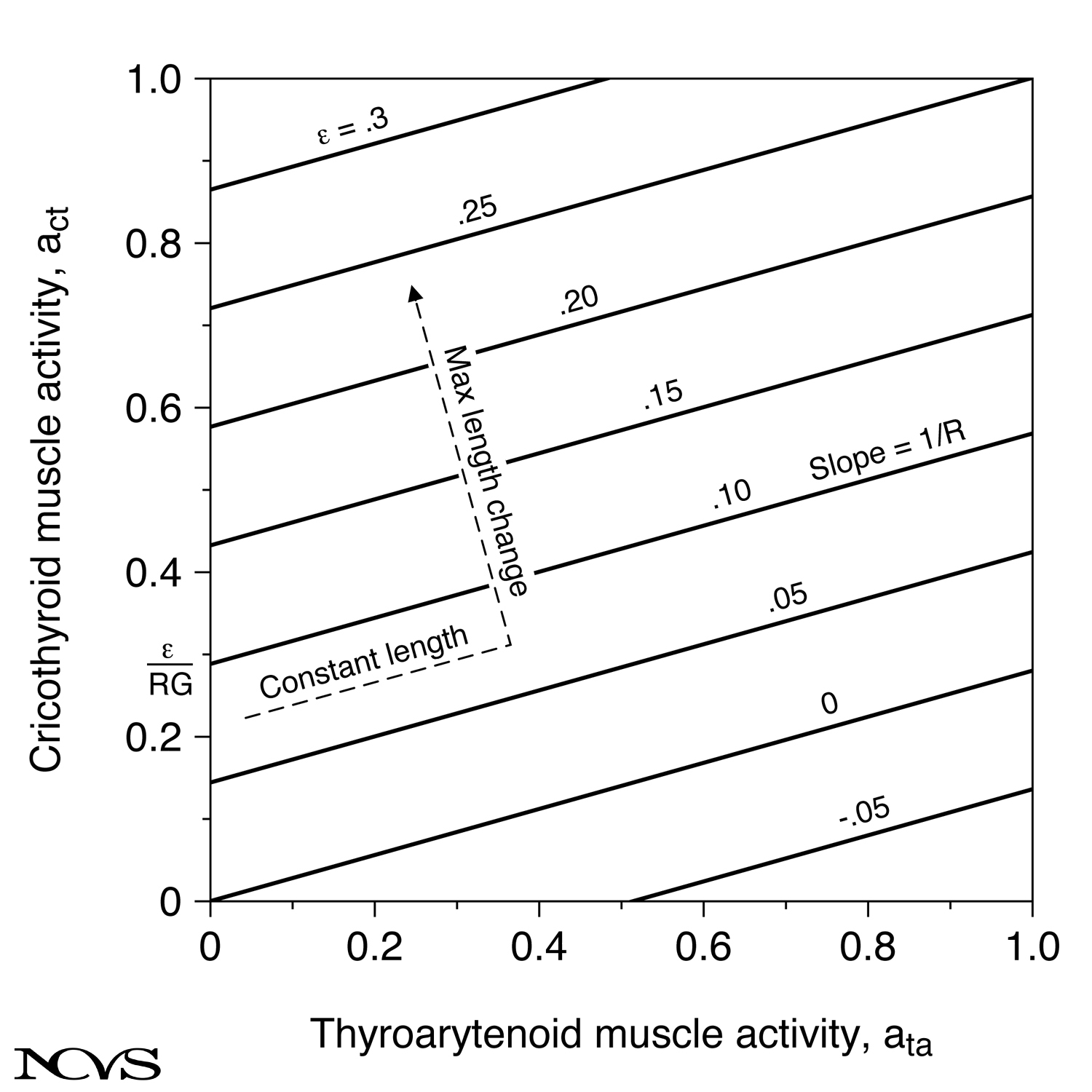 Figure 8.3: Muscle activation plot (MAP) with isometric lines.
A powerful visualization tool is the Muscle Activation Plot:
Axes and Representation
- Vertical axis: Normalized CT activity (αCT)
- Horizontal axis: Normalized TA activity (αTA)
- Sloping lines: Constant elongation (isometric lines)
- Line slope: 1/R (inverse of torque ratio)
Isometric Lines
Lines of constant elongation have slope = 1/R:
Shallow Slope (Small 1/R, Large R)
- CT more effective at changing length than TA
- Small CT change = large TA change for same length
- CT mechanically advantaged
- Typical of human larynx
Steep Slope (Large 1/R, Small R)
- TA more effective at changing length than CT
- Would indicate TA mechanical advantage
- Not typical of normal larynges
Intercepts and Spacing
Vertical Axis Intercept
- Each line intercepts at E/(RG), where E is the normalized elongation (strain)
- Closer spacing indicates larger gain factor
- Wider spacing means limited elongation range
- Individual variation affects vocal capability
Line Spacing
- Uniform spacing only if G constant
- In reality, spacing varies with configuration
- Reflects nonlinear geometric effects
- Closer at longer lengths typically
Isometric Processes
A key concept is the isometric process—muscle activity changes with constant geometry:
Definition
- Change in muscle activities along isometric line
- No net movement results
- Both CT and TA can increase or decrease
- Opposing forces balance
Mechanical Analogy
- Similar to pressing hands together (isometric exercise)
- Large forces develop with no movement
- Muscle tension increases dramatically
- Internal stress increases in tissue
Relevance to Phonation
- Important for F₀ control at constant length
- Enables stiffness change without length change
- Allows register transitions with minimal length change
- Key to advanced vocal technique
Maximum Length Change Trajectory
The path perpendicular to isometric lines represents maximum length change:
Optimal Control Strategy
- Direction of maximum Δℓ per unit muscle activity change
- Slope = -R (negative of torque ratio)
- Requires decreasing αTA while increasing αCT
- Most efficient pitch increase strategy
Practical Implementation
- Vocalist gradually releases TA while engaging CT
- Achieves maximum elongation with minimum effort
- Reduces unnecessary muscle tension
- Often taught in advanced vocal pedagogy
The Dashed Trajectory (Figure 8.3)
The figure shows a typical vocalist’s path:
- Initial phase: Nearly isometric (horizontal movement)
- Both muscles activate to stabilize larynx
- Little length change initially
- Transition: Direction changes toward perpendicular
- TA begins to release
- CT continues to increase
- Final phase: Near-perpendicular direction
- Maximum length increase
- Optimal F₀ rise
Individual Variation
Significant individual differences exist in elongation mechanics:
Anatomical Variation
- Cricothyroid joint morphology varies
- Some joints allow more translation than others
- Lever arm lengths differ
- Muscle size and attachment points vary
Functional Consequences
- Some individuals elongate easily (large G)
- Others have limited elongation range (small G)
- Mechanical advantage (R) affects control strategy
- Training can optimize use of available range but cannot change anatomy
Clinical Implications
- Voice therapy must account for individual mechanics
- Some pitch goals may be anatomically unrealistic
- Understanding limitations prevents frustration
- Alternative strategies may be needed for limited elongation
Extrinsic Muscle Contributions
While the model focuses on intrinsic muscles, extrinsic laryngeal muscles also contribute:
Sternothyroid
- Pulls larynx downward
- May increase effective elongation through external traction
- Could supplement cricothyroid action
- More important at pitch extremes
Other Strap Muscles
- Stabilize laryngeal framework
- Provide platform for intrinsic muscle action
- May assist or oppose elongation
- Individual strategies vary
Summary
Vocal fold elongation results from complex three-dimensional movements of laryngeal cartilages, primarily controlled by the cricothyroid and thyroarytenoid muscles. The CT muscle rotates and translates the thyroid cartilage relative to the cricoid, while the TA opposes this action. The net elongation depends on the difference between CT and TA activities, scaled by geometric and mechanical factors unique to each individual.
The Muscle Activation Plot provides a powerful framework for visualizing how different combinations of CT and TA activity produce elongation. Isometric lines represent constant length, while perpendicular paths represent maximum length change. Understanding these mechanics is essential for vocal pedagogy, voice therapy, and prediction of F₀ control capabilities.
Key Takeaways
- ✅ Vocal fold elongation occurs through rotation and translation at the cricothyroid joint
- ✅ Cricothyroid muscle has two parts: pars recta (rotation) and pars obliqua (translation)
- ✅ Thyroarytenoid muscle opposes CT action, creating differential control
- ✅ Net elongation depends on difference between CT and TA activities, not absolute levels
- ✅ Torque ratio (R) describes mechanical advantage of CT over TA
- ✅ Gain factor (G) determines range of achievable elongation
- ✅ Isometric lines on MAP represent constant vocal fold length
- ✅ Maximum length change occurs perpendicular to isometric lines
- ✅ Individual anatomical variation significantly affects elongation capability
Related Topics
- Analogies with Vibrating Strings and Ribbons
- Quantitative F₀ Analysis for the Cover Model
- Muscle Activation Plot for the Body-Cover Model
Further Reading
- Titze, I. R., Jiang, J. J., & Druker, D. G. (1988). Preliminaries to the body-cover theory of pitch control. Journal of Voice, 1, 314-319.
- Honda, K. (1983). Relationship between pitch control and vowel articulation. In I. R. Titze & R. C. Scherer (Eds.), Vocal Fold Physiology: Biomechanics, Acoustics, and Phonatory Control (pp. 286-297). Denver Center for the Performing Arts.
- van den Berg, J. (1958). Myoelastic-aerodynamic theory of voice production. Journal of Speech and Hearing Research, 1, 227-244.