Hard-Tissue Morphology
The larynx’s remarkable mobility within the neck is made possible by its primary structural component: cartilage rather than bone. This cartilaginous framework allows the larynx to move vertically several centimeters during swallowing and yawning, shift forward when airflow is needed, and adjust position for optimal voice production. Understanding why the larynx evolved as a cartilaginous rather than bony structure reveals fundamental principles about the balance between stability and mobility in biological systems.
Laryngeal Mobility and Functional Demands
The position of the larynx within the neck varies considerably depending on physiological demands. During swallowing, a group of sphincter muscles forces the larynx upward so the airway can be sealed, preventing food or liquid from entering the trachea. During yawning, the pharynx expands and the larynx is depressed to widen the airway for maximum inhalation. These maneuvers can move the larynx vertically by several centimeters.
The larynx can also move forward when greater-than-normal airflow is needed or when a bolus of food enters the pharynx. This mobility enables the larynx to perform multiple functions: airway protection, breathing, and phonation. Each function requires different laryngeal positions and configurations.
Why Cartilage Instead of Bone?
The need for mobility of the entire laryngeal structure precludes rigid bony attachments to the skeleton. Bone would create fixed anchor points that limit movement. Cartilage, by contrast, provides structural support while maintaining flexibility. This explains why most of the laryngeal framework is composed of cartilage.
Only one bone—the hyoid bone—is in the vicinity of the larynx, and even this bone “floats” with respect to the skeleton, having no direct bony connections. The hyoid serves as a mobile anchor point for muscles and ligaments but does not restrict laryngeal movement.
Cartilage Composition and Properties
Laryngeal cartilages are composed primarily of hyaline cartilage (in the thyroid, cricoid, and most of the arytenoid cartilages) and elastic cartilage (in the epiglottis, cuneiform, and corniculate cartilages). These two types have distinct properties:
Hyaline Cartilage:
- Provides firm structural support
- Contains collagen fibers embedded in a gel-like matrix
- Capable of calcification and ossification with age
- Found in the major laryngeal cartilages (thyroid, cricoid, arytenoid)
Elastic Cartilage:
- More flexible and deformable than hyaline cartilage
- Contains abundant elastin fibers for rebound resilience
- Does not ossify with age
- Found in cartilages requiring repeated bending (epiglottis)
Ossification Patterns
With aging, hyaline cartilages undergo progressive ossification—the replacement of cartilage with bone tissue. This process typically begins in adulthood and continues throughout life. Ossification patterns vary considerably among individuals but generally follow predictable sequences.
The thyroid cartilage typically shows ossification beginning in the inferior cornua and progressing through the laminae. The cricoid cartilage often ossifies from posterior to anterior. The arytenoid cartilages may show partial ossification, particularly at the muscular processes.
Ossification has important implications for vocal function. Increased stiffness from calcification can affect cartilage mobility at joints, potentially influencing pitch range and vocal flexibility. However, the relationship between ossification and voice changes is complex and not fully understood.
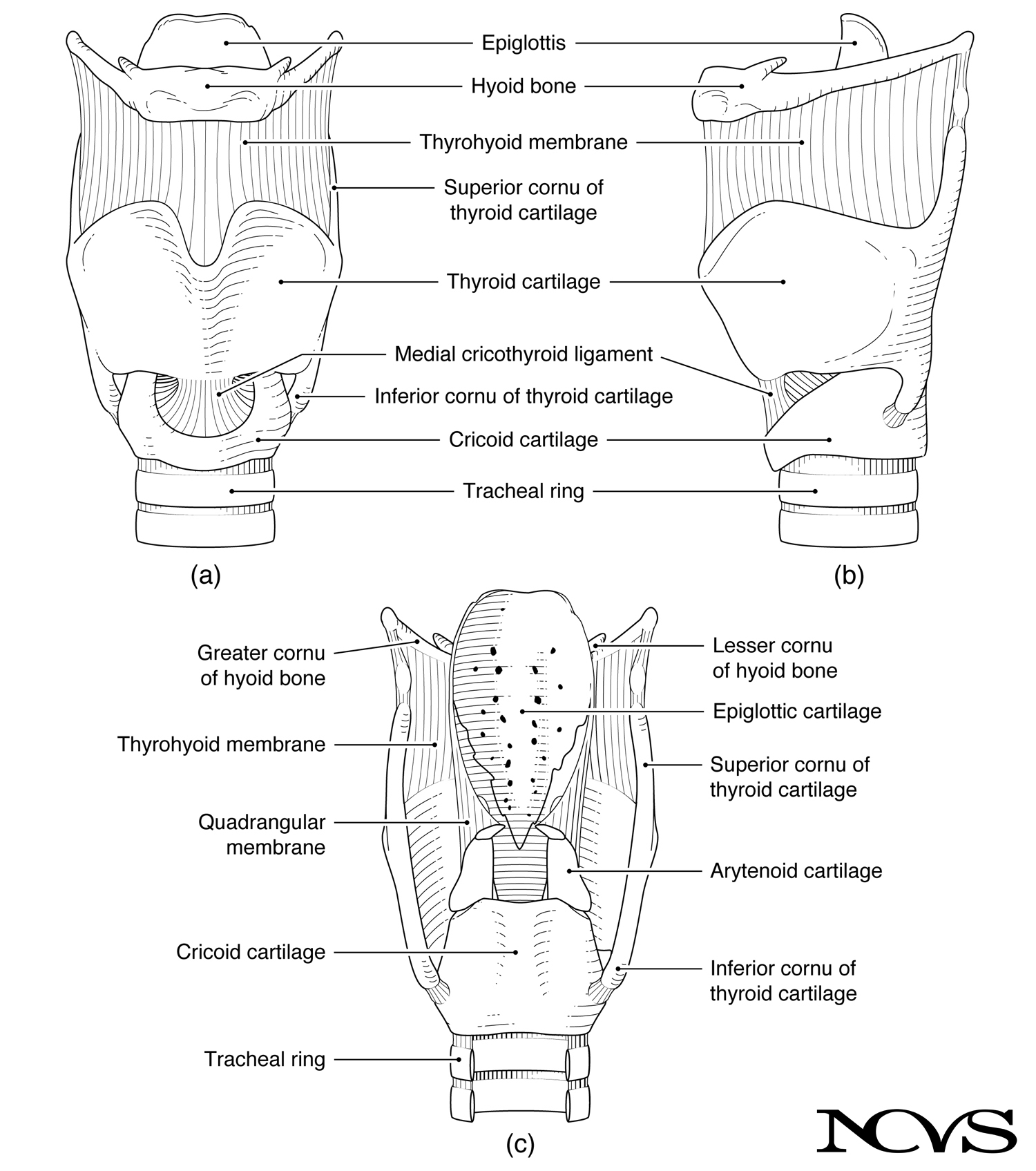 Figure 1.3: Framework of the larynx showing major cartilages and their interconnections. The cartilaginous structure allows mobility while providing necessary support.
Ligaments and Membranes
Connecting the cartilages are various ligaments (connecting bone to bone or cartilage to cartilage) and membranes (sheet-like connective tissue). These soft connective tissues maintain structural relationships while permitting movement.
Key connecting structures include:
- Thyrohyoid membrane: connects the thyroid cartilage to the hyoid bone
- Cricothyroid membrane (also called the medial cricothyroid ligament): connects the cricoid and thyroid cartilages anteriorly
- Cricoarytenoid joints: synovial joints allowing arytenoid rotation and gliding
- Cricothyroid joints: paired joints enabling thyroid cartilage rotation relative to the cricoid
- Vocal ligament: the thickened free edge of the conus elasticus, forming the medial edge of the vocal fold
These connective structures enable coordinated movement of the laryngeal cartilages while maintaining the integrity of the framework.
Summary
The larynx’s cartilaginous framework represents an elegant solution to competing functional demands. Cartilage provides sufficient structural support for muscle attachment and airway maintenance while allowing the mobility essential for swallowing, breathing, and phonation. The hyoid bone, though osseous, floats freely to support this mobile system.
The composition of laryngeal cartilages—hyaline cartilage in major structural components and elastic cartilage in flexible elements—reflects their specific functional roles. Age-related ossification demonstrates how laryngeal structures change over the lifespan, with potential implications for voice production.
Understanding hard-tissue morphology provides context for how muscles, acting on this cartilaginous framework, generate the fine motor control necessary for vocal fold positioning and tension regulation.
Key Takeaways
- ✅ The larynx is primarily cartilaginous, enabling mobility for swallowing, breathing, and phonation
- ✅ Hyaline cartilage provides structural support in major laryngeal cartilages
- ✅ Elastic cartilage in the epiglottis and other flexible structures resists ossification
- ✅ Age-related ossification progressively stiffens laryngeal cartilages
- ✅ The hyoid bone floats relative to the skeleton, supporting laryngeal mobility
- ✅ Ligaments and membranes interconnect cartilages while permitting movement
Related Topics
Further Reading
- Romanes, G. J. (1964). Cunningham’s textbook of anatomy. Oxford University Press.
- Kahane, J. (1978). A morphological study of the human prepubertal and pubertal larynx. American Journal of Anatomy, 151, 11-20.
- Maue, W. M. (1970). Cartilages, ligaments and articulations of the adult human larynx. Dissertation, University of Pittsburgh.