The Hyoid Bone
The hyoid bone occupies a unique position in human anatomy: it is the only bone in the body that does not articulate directly with any other bone. This horseshoe-shaped structure “floats” in the anterior neck, suspended by muscles and ligaments above and below. Despite not being part of the larynx proper, the hyoid bone plays a critical role in laryngeal function, serving as a mobile anchor point that coordinates movements of the tongue, jaw, larynx, and pharynx.
Structural Anatomy
The hyoid bone consists of a body (corpus) and two pairs of projections called horns or cornua:
Body: The central, horizontally oriented portion that forms the anterior curve of the horseshoe. It is broader and more substantial in males than females.
Greater Cornua (greater horns): Two longer, backward-projecting processes that extend from each end of the body. These provide attachment points for numerous muscles and ligaments, including the middle pharyngeal constrictor muscle and the thyrohyoid membrane.
Lesser Cornua (lesser horns): Two smaller, upward-projecting processes located at the junction between the body and greater cornua. These are connected to the styloid process of the temporal bone via the stylohyoid ligament, linking the hyoid to the skull base.
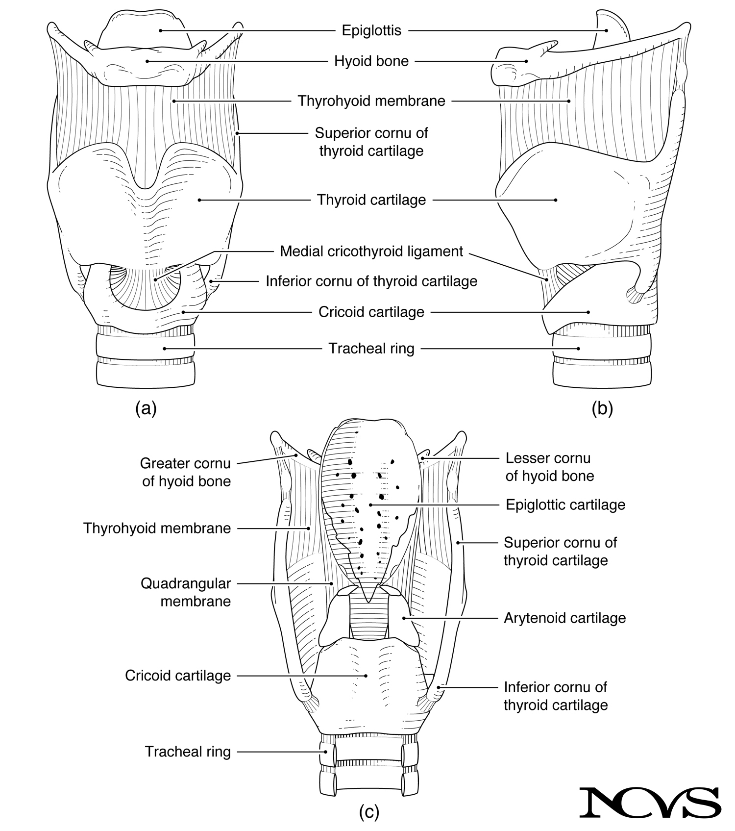 Figure 1.3: The hyoid bone shown in relation to laryngeal cartilages in anterior, lateral, and posterior views.
The hyoid can be viewed from anterior, lateral, and posterior aspects (Figure 1.3). Its position partially surrounds the tip of the epiglottis, with the epiglottis attaching to its posterior surface via the hyoepiglottic ligament (see Figure 1.8b in the Laryngeal Cartilages topic).
Connections to the Larynx
Although the hyoid is not part of the laryngeal framework, it connects intimately to laryngeal structures:
Thyrohyoid Membrane: A broad, fibro-elastic membrane connecting the superior border of the thyroid cartilage to the inferior border of the hyoid body and greater cornua. This membrane contains a thickened central portion (the median thyrohyoid ligament) and thickened lateral portions (the lateral thyrohyoid ligaments) that connect to the superior cornua of the thyroid cartilage.
Thyrohyoid Muscle: This paired muscle runs vertically from the oblique line of the thyroid lamina to the inferior border of the hyoid body and greater cornua. When contracted, it can either elevate the larynx (if the hyoid is fixed) or depress the hyoid (if the larynx is fixed).
The vertical distance between the hyoid bone and thyroid cartilage changes during phonation, swallowing, and other laryngeal functions. This adjustment affects the configuration of the pharyngeal space and can influence vocal tract resonance.
The Hyoid as a “Hitching Post”
One might think of the hyoid bone as the hitching post for tissue connections needed to form an approximate 90° bend in the airway from the mouth to the pharynx. Multiple muscle groups attach to the hyoid:
From above (suprahyoid muscles): Connect the hyoid to the skull, jaw, and tongue From below (infrahyoid muscles): Connect the hyoid to the larynx and sternum From behind (pharyngeal muscles): Connect the hyoid to the pharyngeal wall
This arrangement creates a three-dimensional suspension system. The hyoid’s position is determined by the balance of forces from these various muscle groups, allowing for coordinated movement during breathing, swallowing, and speaking.
Protective Function
The hyoid bone helps protect soft tissues in the upper larynx and pharynx against injury from blows to the neck. Its horseshoe shape distributes forces around critical structures rather than transmitting impact directly to the laryngeal cartilages or airway. While the hyoid can be fractured (particularly in cases of strangulation), its presence provides some mechanical protection for underlying structures.
Hyoid Position and Voice Production
Hyoid position influences the configuration of the pharynx and thus affects vocal tract resonance. Laryngeal height—the vertical position of the larynx in the neck—is partly determined by the relationship between the hyoid and the thyroid cartilage.
Voice teachers and clinicians recognize that hyoid position affects voice quality:
Elevated larynx/hyoid: Generally associated with pharyngeal constriction and a more “tight” or “bright” vocal quality. Common in untrained singers at high pitches or when experiencing vocal tension.
Neutral larynx/hyoid: Typically preferred for classical singing and speech, providing balanced resonance without excessive tension.
Lowered larynx/hyoid: Often cultivated in operatic singing to create a larger pharyngeal space and “darker” vocal timbre. However, excessive depression can create tension and instability.
The extrinsic laryngeal muscles that attach to the hyoid control these postural adjustments. Understanding hyoid mobility is essential for voice pedagogy and therapy addressing issues of laryngeal tension.
Palpation and Clinical Examination
The hyoid bone is readily palpable in the anterior neck, located approximately at the level of the third cervical vertebra (C3), above the thyroid cartilage. To locate it:
- Place fingers at the midline of the upper neck, just below the chin
- Press gently backward and downward
- The hard, mobile structure felt is the hyoid body
- Ask the patient to swallow—the hyoid elevates noticeably during the swallow
Palpation of the hyoid can provide clinical information about:
- Hyoid mobility during swallowing
- Laryngeal elevation patterns
- Muscle tension in the anterior neck
- Presence of masses or abnormalities in the region
Reduced hyoid excursion during swallowing may indicate weakness of suprahyoid muscles and can be associated with dysphagia (swallowing difficulty).
Summary
The hyoid bone occupies a central position in the biomechanics of the upper airway and neck. As the only bone in the region, floating without direct skeletal connections, it serves as a mobile anchor that coordinates movements of multiple muscle groups. Its connections to the larynx via the thyrohyoid membrane and thyrohyoid muscle make it integral to laryngeal function, despite not being a laryngeal cartilage itself.
The hyoid’s role as a “hitching post” for the approximately 90° airway bend from mouth to pharynx demonstrates how anatomical structures serve multiple functions. Its position influences pharyngeal shape and thus vocal tract resonance, making hyoid mobility an important consideration in voice production. Clinically, hyoid palpation and observation of its movement during swallowing provide valuable diagnostic information.
Key Takeaways
- ✅ The hyoid is the only bone that does not articulate with any other bone, floating via muscular and ligamentous connections
- ✅ It consists of a body, paired greater cornua (horns), and paired lesser cornua
- ✅ The thyrohyoid membrane connects the hyoid to the thyroid cartilage, coordinating their movement
- ✅ The hyoid serves as an anchor point for suprahyoid muscles (above), infrahyoid muscles (below), and pharyngeal muscles (posterior)
- ✅ Hyoid position influences laryngeal height and pharyngeal configuration, affecting vocal resonance
- ✅ The hyoid is readily palpable and its movement during swallowing can be clinically assessed
Related Topics
Further Reading
- Palmer, J. M. (1984). Anatomy for speech and hearing (3rd ed.). New York: Harper & Row.
- Kahane, J., & Folkins, J. (1984). Atlas of speech and hearing anatomy. Columbus, OH: Charles E. Merrill Publishing.
- Zemlin, W. R. (1964). Speech and hearing science: Anatomy and physiology. Champaign, IL: Stipes Publishing Co.