Morphology of Vocal Fold Soft Tissue
The vocal folds are among the most biomechanically sophisticated structures in the human body. Their ability to vibrate hundreds of times per second, sustain this vibration for extended periods, withstand the mechanical stresses of collision, and modulate precisely in response to neuromuscular commands depends critically on their microstructural organization. Understanding the layered architecture of vocal fold tissue is essential for comprehending how voice is produced and how pathological changes affect vocal function.
The Vocal Tract and Glottal Position
Before examining vocal fold tissue in detail, it is important to understand their position within the larger vocal tract. Figure 1.12 shows a coronal (frontal) section through the larynx, revealing the airway between the trachea and the tip of the epiglottis. The vocal folds are located at the narrowest portion of this airway, positioned horizontally across the laryngeal opening.
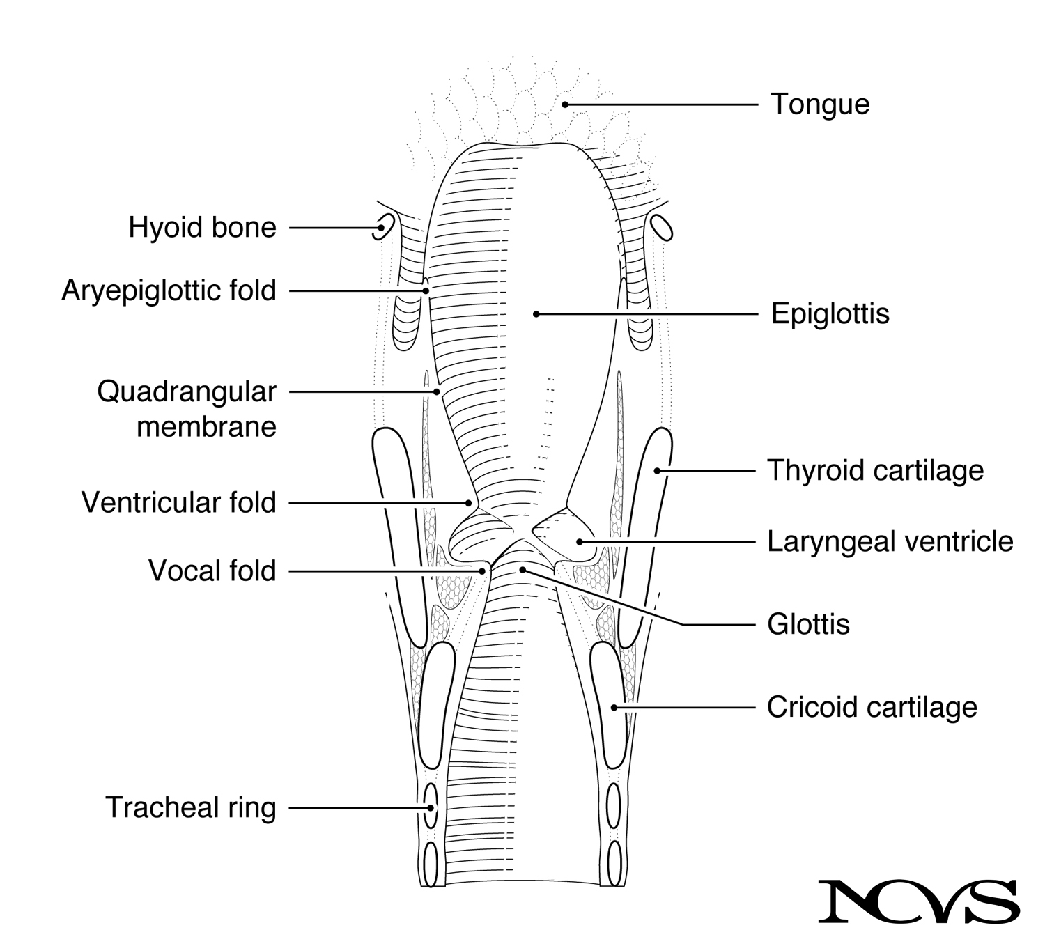 Figure 1.12: Coronal section through the larynx showing the laryngeal airway. Note the vocal folds at the narrowest point, with the laryngeal ventricle (sinus of Morgagni) above them.
Above the vocal folds are the ventricular folds (also called false vocal folds), which are thicker, more bulky folds that do not normally participate in phonation but serve protective functions. The space between the vocal folds and ventricular folds is the laryngeal ventricle (also known as the sinus of Morgagni).
Above the ventricular folds are the aryepiglottic folds, which together with the epiglottis produce a narrowing in the collar of the larynx (see Figure 1.8b). In combination, the vocal folds, ventricular folds, aryepiglottic folds, and quadrangular membrane (a sheet of connective tissue) constitute a system of folds that can seal off the laryngeal airway rapidly and completely when appropriate muscles are activated.
The Layered Structure of the Vocal Fold
The vocal fold is not a homogeneous structure but rather a highly organized system of layers, each with distinct mechanical properties. This layering enables the complex vibratory patterns necessary for voice production.
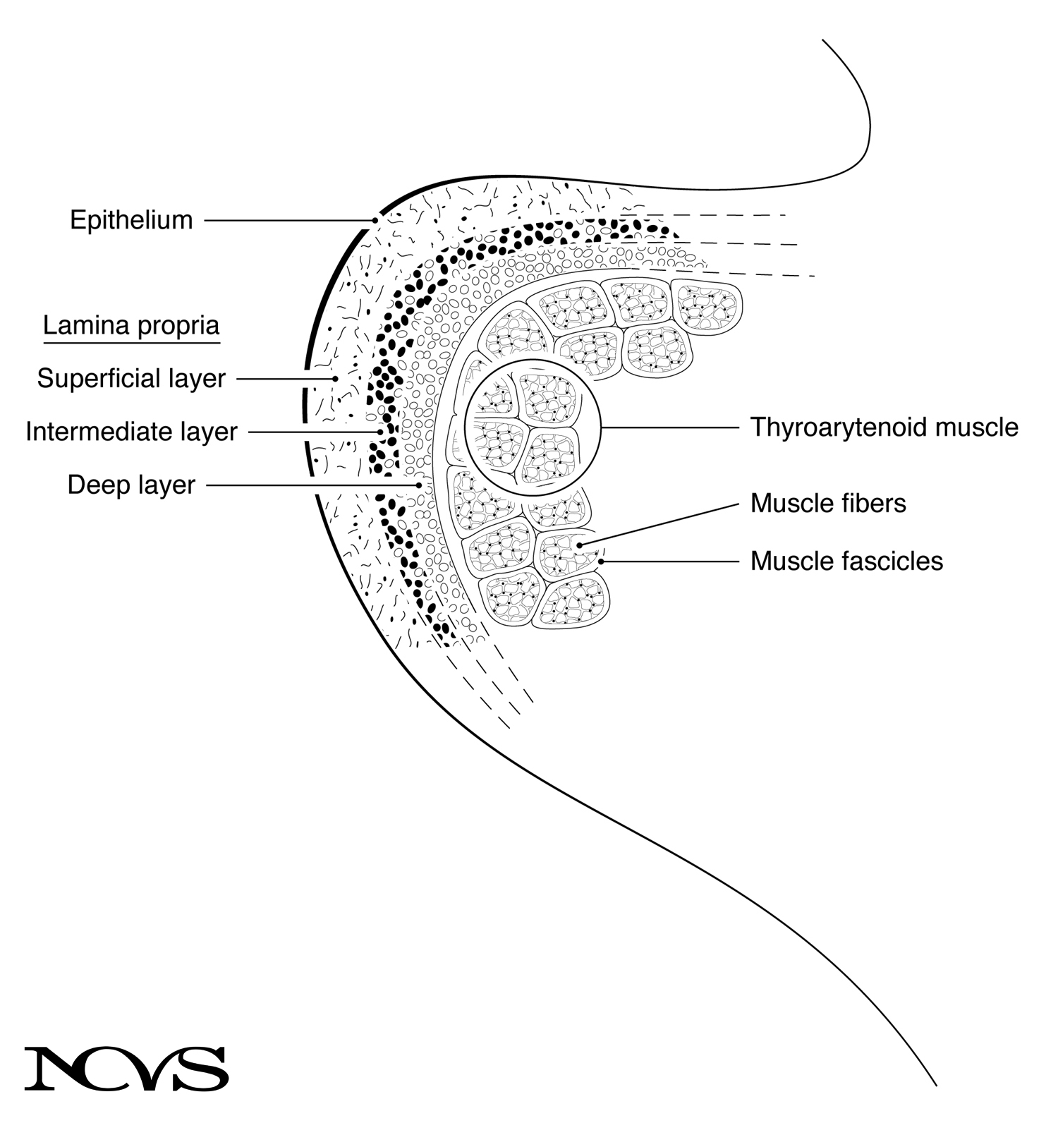 Figure 1.13: Schematic of a coronal section through the right vocal fold, showing tissue layers from superficial to deep.
The Epithelium
The outermost layer is a thin skin made up of stratified squamous (layered and scalelike) epithelium. This layer is approximately 0.05 to 0.10 mm thick—about the thickness of a sheet of paper. The epithelium encapsulates the softer, fluid-filled tissues beneath, somewhat like a balloon filled with water.
The epithelial layer serves several critical functions:
Barrier Protection: Creates a sealed surface that prevents fluid loss and protects underlying tissues from trauma during vocal fold collision.
Smooth Gliding Surface: The epithelium must allow the overlying mucus layer to move freely during the mucosal wave (the wavelike motion of the vocal fold surface during vibration).
Structural Integrity: Despite being thin, the epithelium must withstand the repetitive mechanical stresses of phonation without tearing or developing lesions.
The vocal fold epithelium is non-keratinized (unlike skin), which keeps it moist and pliable. However, chronic irritation (from smoking, reflux, or vocal trauma) can cause epithelial thickening or keratinization, altering the mechanical properties of the vocal fold surface.
The Lamina Propria
Beneath the epithelium lies the lamina propria, a layered system of nonmuscular tissues between the epithelium and muscle. The lamina propria is conveniently divided into three layers with distinct compositions and mechanical properties:
Superficial Layer of the Lamina Propria (Reinke’s Space)
The superficial layer consists primarily of loosely organized elastin fibers surrounded by interstitial fluids. Elastin fibers are made of a special type of protein structure that allows for ample elongation, like a rubber band.
This layer is approximately 0.5 mm thick in the middle of the vocal fold and is sometimes called Reinke’s space, referring to the potential space that can fill with fluid in pathological conditions (Reinke’s edema).
Mechanical Properties:
- Very low viscosity and elasticity compared to deeper layers
- Allows large-amplitude deformation with minimal resistance
- Facilitates the mucosal wave by decoupling the epithelium from stiffer underlying structures
The superficial layer’s loose organization is critical for normal phonation. When this layer becomes fibrotic (filled with scar tissue) or edematous (filled with excess fluid), vocal fold vibration is severely impaired.
Intermediate Layer of the Lamina Propria
The intermediate layer is also made of elastin fibers, but they are more densely packed and oriented primarily in the anterior-posterior (longitudinal) direction (shown as filled dots in Figure 1.13). This orientation reflects the direction of stress during phonation—elastin fibers align with the primary direction of tissue deformation.
Interspersed with the elastin are collagen fibers (unfilled dots in Figure 1.13). Collagen fibers have a protein structure that, like a cotton thread, makes them nearly inextensible. The presence of collagen begins to add stiffness to this layer compared to the superficial layer.
The intermediate layer is approximately 1 to 2 mm thick. Together with the deep layer, it forms what is functionally known as the vocal ligament.
Deep Layer of the Lamina Propria
The deep layer is made primarily of collagen fibers, giving it much greater stiffness than the superficial or intermediate layers. These fibers also run parallel along the anterior-posterior direction.
Like a cotton thread, collagen fibers are nearly inextensible. The deep layer’s high collagen content makes it the stiffest component of the lamina propria, providing structural support while still allowing controlled deformation.
The fibers in the deep layer also run parallel along the anterior-posterior direction, and together with the intermediate layer, this portion is approximately 1 to 2 mm thick.
The Thyroarytenoid Muscle
The thyroarytenoid muscle, discussed previously as an intrinsic laryngeal muscle, lies beneath the lamina propria. This is the major portion of the vocal fold mass, approximately 7 to 8 mm thick.
Muscle is usually organized in fascicles (bundles), as shown in Figure 1.13, although the divisions are not always as clear to the eye as the schematic suggests. The muscle fibers run in the anterior-posterior direction, from the thyroid cartilage to the arytenoid cartilage.
Mechanical Properties:
- Can actively contract and generate tension
- Stiffness varies with activation level
- Forms the “body” of the vocal fold in biomechanical models
The muscle’s ability to stiffen through contraction provides active control over vocal fold mechanical properties, enabling adjustments to fundamental frequency and vocal intensity.
Labeling Schemes for Tissue Layers
Different labeling schemes have been used to group the tissue layers, depending on the physiological or biomechanical concept being described. Understanding these schemes is important for reading the voice science literature.
Three-Layer Scheme
The traditional anatomical scheme divides the vocal fold into:
Mucosa: Epithelium + superficial layer of lamina propria
Ligament: Intermediate layer + deep layer of lamina propria
Muscle: Thyroarytenoid muscle
This scheme emphasizes the distinct tissue types: mucosa (epithelial and loose connective tissue), ligament (dense connective tissue), and muscle (contractile tissue).
Five-Layer Scheme
The microanatomical scheme divides the vocal fold into five distinct layers:
- Epithelium
- Superficial layer of lamina propria
- Intermediate layer of lamina propria
- Deep layer of lamina propria
- Muscle
This scheme, popularized by Hirano and colleagues, provides the most detailed description of vocal fold microstructure and is particularly useful for understanding the biomechanics of phonation and the effects of vocal pathology.
Two-Layer Scheme (Cover-Body Theory)
The biomechanical scheme divides the vocal fold into two functional units:
Cover: Epithelium + superficial layer + intermediate layer of lamina propria
Body: Deep layer of lamina propria + muscle
This scheme underlies the cover-body theory of vocal fold vibration, which describes how the cover can vibrate somewhat independently of the body during phonation. The cover’s relatively loose coupling to the body enables the mucosal wave—the wavelike rippling of the vocal fold surface that is characteristic of normal phonation.
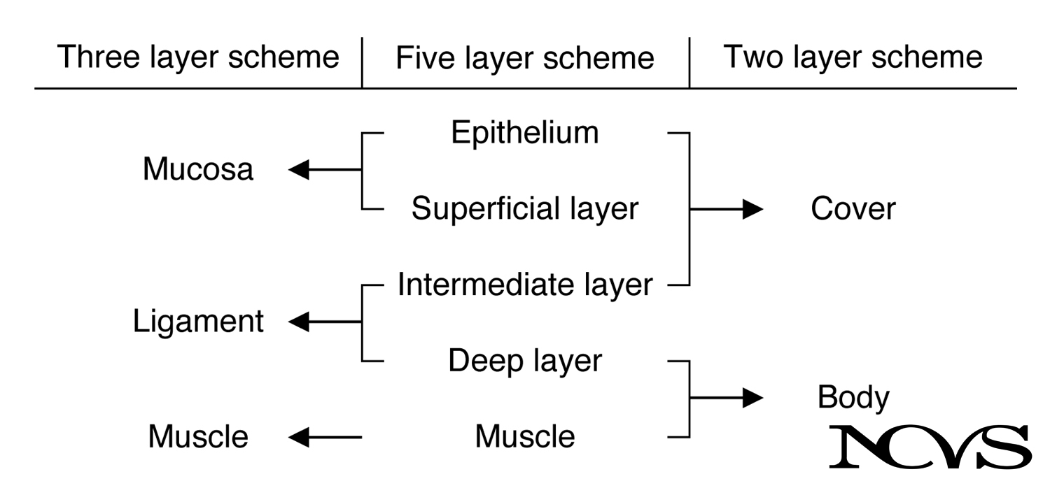 Figure 1.14: Three different schemes used for labeling the layered structure of the vocal fold.
Tissue Composition and Fiber Orientation
The organization of elastin and collagen fibers within the lamina propria reflects the biomechanical demands placed on vocal fold tissue.
Elastin Fibers:
- Predominant in superficial and intermediate layers
- Provide elastic recoil—the tissue returns to its original shape after deformation
- Allow large deformations with relatively low force
- Oriented somewhat randomly in superficial layer; more longitudinally oriented in intermediate layer
Collagen Fibers:
- Predominant in intermediate and deep layers
- Provide tensile strength—resist elongation
- Limit maximum tissue deformation
- Oriented longitudinally (anterior-posterior direction)
The longitudinal orientation of fibers in the intermediate and deep layers is particularly important. During phonation, the vocal folds are stretched longitudinally (lengthened), creating tension along their length. The anterior-posterior fiber orientation provides maximum resistance to this stretch, allowing the vocal folds to sustain high tensions without excessive elongation.
Vocal Ligament Thickening at Endpoints
The vocal fold is not uniformly thick along its length. Histological examination reveals that the vocal ligament is thicker at the endpoints—near the anterior commissure (where the vocal folds attach to the thyroid cartilage) and at the vocal processes of the arytenoid cartilages.
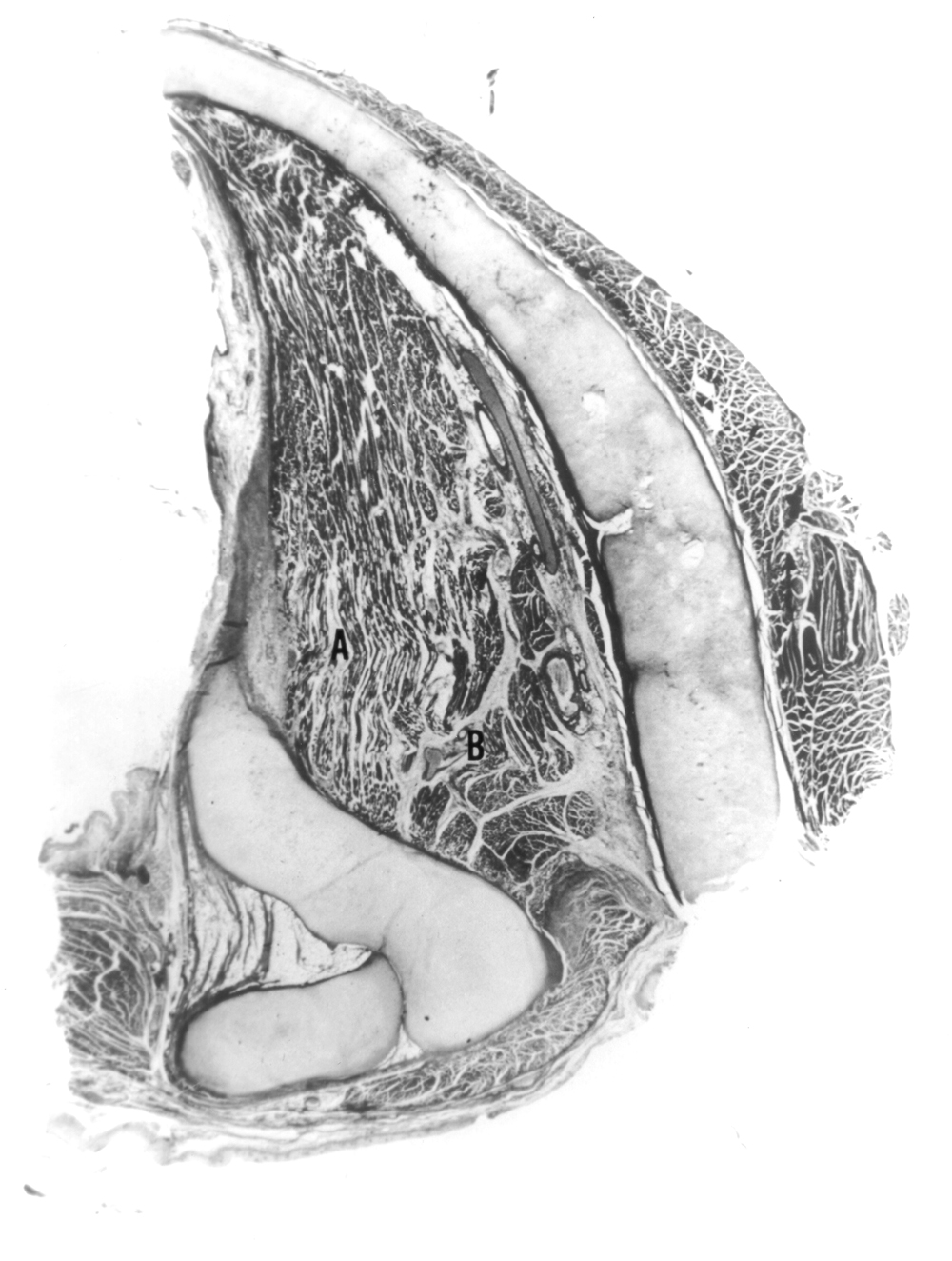 Figure 1.15: Histological section through the right vocal fold, revealing the thickening of the vocal ligament at the end points (dark layer near left edge). This section was taken at the level of the vocal process. From H. von Leden, Archives of Otolaryngology, 74 (1961), 660-676. Copyright 1961, American Medical Association.
This thickening likely represents an evolutionary adaptation to protect against stress concentration at the endpoints. During phonation, the vocal folds are under longitudinal tension, and mechanical analysis shows that stresses are highest at the attachment points. The reinforced ligament at these locations helps prevent tissue damage from these repetitive high stresses.
Additionally, the mucosa is thicker in the middle of the vocal fold, where most of the “head-on” collision occurs between the left and right vocal folds during the closing phase of vibration. This thickening may provide a cushion (shock absorber) for the ligament, protecting it from impact forces.
Species Differences: Comparison with Canine Larynx
The canine (dog) larynx is frequently used as an animal model for studying human voice production. However, there are important structural differences between canine and human vocal folds.
Comparison of canine vocal fold tissue with human vocal fold tissue reveals that the canine does not have a well-established ligament (intermediate and deep layers of lamina propria). The canine’s superficial layer is thicker (approximately 2 to 3 mm, compared to 0.5 mm in humans), and the intermediate and deep layers are largely nonexistent.
Functional Implications:
The significance of this difference can only be speculated, but one hypothesis is that the human vocal ligament is crucial for sustaining high pitches. High-pitched phonation requires large longitudinal tensions in the collagenous fibers of the ligament. The mucosa can remain relatively lax for ease of vibration while the ligament sustains the tension.
Dogs can initiate phonation but seem less able to sustain it, especially at high pitches. The thicker mucosa of the canine vocal fold is appropriate for barking—bursts of phonation with rapid onset. For steady phonation at varied pitches, a more differentiated structure with a distinct vocal ligament may be advantageous.
This comparison highlights how vocal fold structure has adapted to meet the specific phonatory demands of different species.
Clinical Implications of Layered Structure
Understanding vocal fold microstructure is essential for comprehending voice pathology and treatment:
Vocal Fold Lesions
Nodules: Benign masses that typically form at the junction of the anterior and middle thirds of the vocal folds (the point of maximum collision force). They involve thickening of the epithelium and superficial lamina propria.
Polyps: Often develop in Reinke’s space (superficial lamina propria), filled with edematous tissue or blood vessels. They disrupt the mucosal wave by adding mass to the cover.
Cysts: Fluid-filled or mucus-filled lesions, often in the superficial lamina propria, that can be congenital or acquired. They restrict vibration of the affected vocal fold.
Scarring and Fibrosis
Scar tissue replaces normal lamina propria architecture with disorganized collagen, eliminating the layered structure. The superficial layer becomes stiff, severely impairing the mucosal wave. Vocal fold scarring is one of the most difficult voice problems to treat because the tissue cannot regenerate its original layered architecture.
Sulcus Vocalis
A sulcus is a groove or depression along the vocal fold edge, often involving fusion of epithelium to deeper layers or absence of the superficial lamina propria. This condition restricts vibration and causes a weak, breathy voice.
Reinke’s Edema
Chronic swelling of Reinke’s space (the superficial lamina propria) causes the vocal fold to become “waterlogged” and floppy. This typically results in a very low-pitched, rough voice. The condition is strongly associated with smoking and chronic laryngeal irritation.
Summary
The vocal fold is a marvel of biological engineering, with a precisely layered microstructure that enables both flexibility and strength. The epithelium provides a protective barrier, the superficial lamina propria allows large-amplitude vibration with minimal resistance, the intermediate and deep layers (vocal ligament) sustain longitudinal tension while limiting deformation, and the thyroarytenoid muscle actively controls stiffness and position.
The organization of elastin and collagen fibers reflects the biomechanical demands of phonation, with fiber orientations and concentrations optimized for the specific stress patterns experienced during voice production. The vocal ligament’s reinforcement at endpoints and the mucosa’s thickening at the point of maximum collision demonstrate how tissue structure has evolved to withstand the repetitive mechanical stresses of phonation.
Different labeling schemes—the three-layer, five-layer, and two-layer (cover-body) models—provide complementary perspectives on vocal fold structure. The five-layer scheme offers the most detailed anatomical description, while the cover-body model is most useful for understanding phonatory biomechanics.
Understanding this microstructural organization is fundamental for comprehending normal voice production, interpreting laryngeal imaging, diagnosing voice disorders, and planning surgical or behavioral interventions.
Key Takeaways
- ✅ The vocal fold consists of five distinct layers: epithelium, three layers of lamina propria, and muscle
- ✅ The superficial lamina propria (Reinke’s space) is loosely organized, enabling the mucosal wave
- ✅ The intermediate and deep layers form the vocal ligament, with longitudinally oriented collagen providing tensile strength
- ✅ The cover-body theory divides the vocal fold into a flexible cover and a stiff body, explaining independent surface vibration
- ✅ The vocal ligament is thicker at endpoints to withstand stress concentration; mucosa is thicker at the collision point
- ✅ Disruption of the layered structure (scarring, edema, lesions) severely impairs phonation
Related Topics
Further Reading
- Hirano, M. (1974). Morphological structure of the vocal cord as a vibrator and its variations. Folia Phoniatrica, 26, 89-94.
- Hirano, M. (1977). Structure and vibratory behavior of the vocal folds. In M. Sawashima & F. S. Cooper (Eds.), Dynamic aspects of speech production (pp. 13-30). Tokyo: University of Tokyo Press.
- Hirano, M., Kurita, S., & Nakashima, T. (1981). The structure of the vocal folds. In K. N. Stevens & M. Hirano (Eds.), Vocal fold physiology (pp. 33-41). Tokyo: University of Tokyo Press.
- Gray, S. D., Titze, I. R., & Lusk, R. P. (2000). Electron microscopy of vocal fold tissues. Journal of Voice, 14(1), 95-108.
- Hirano, M., & Kakita, Y. (1985). Cover-body theory of vocal cord vibration. In R. G. Daniloff (Ed.), Speech science (pp. 1-46). San Diego: College Hill Press.