Laryngeal Cartilages
The laryngeal framework consists of nine cartilages: three single (thyroid, cricoid, epiglottis) and three paired (arytenoid, corniculate, cuneiform). These cartilages form the structural skeleton of the larynx, providing attachment points for muscles and ligaments while protecting the airway. Each cartilage has a distinct shape optimized for its specific functions in breathing, swallowing, and voice production.
The Thyroid Cartilage
The thyroid cartilage is the largest of the laryngeal cartilages and forms much of the anterior and lateral walls of the larynx. Its name derives from its shield-like shape (Greek thyreos = shield). This cartilage is made up of two plates, or laminae, that join at the midline anteriorly.
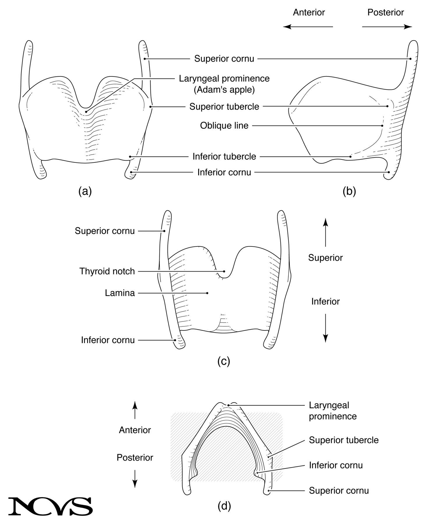 Figure 1.4: The thyroid cartilage shown in (a) anterior view, (b) lateral view, (c) posterior view, and (d) superior view.
Structural Features
The two laminae meet at an angle of approximately 90° to 120° (Figure 1.4d). This angle varies by sex and age: adult males typically have a smaller angle (closer to 90°), resulting in a more prominent laryngeal projection commonly known as the Adam’s apple (laryngeal prominence). Women and children generally have a larger angle (closer to 120°), creating a less pronounced anterior projection.
According to folklore, the first man, Adam, swallowed an apple that became stuck in the larynx, creating a visible protrusion at the front of the neck. While anatomically inaccurate, this cultural story explains the common name for this landmark.
When viewed from the anterior or posterior aspect, a superior thyroid notch is visible at the top where the two laminae meet (Figure 1.4a, c). This notch can easily be palpated on one’s own neck by placing a finger at the midline of the throat and moving upward until a V-shaped depression is felt. A vertical line through this notch would identify the “seam” where the left and right laminae are joined.
Projections and Attachments
From each lamina, two projections arise from the posterior borders of the cartilage:
Superior Cornu (horn): The upward projection connects via the lateral thyrohyoid ligament to the hyoid bone (see Figure 1.3). This connection allows coordinated movement between the thyroid cartilage and hyoid bone.
Inferior Cornu: The downward projection articulates with the cricoid cartilage at the cricothyroid joint (see Figure 1.3). This synovial joint is critical for pitch control, as it allows the thyroid cartilage to rotate relative to the cricoid, thereby elongating or shortening the vocal folds.
Two smaller projections called superior and inferior tubercles (little bumps) constitute the lateral extremities of the thyroid cartilage (Figure 1.4a, b). These tubercles serve as attachment points for muscles and ligaments. An oblique line runs between these tubercles on the external surface of each lamina, providing attachment for the sternothyroid and thyrohyoid muscles.
Clinical Significance
The thyroid cartilage is easily palpable and serves as a key landmark for clinical examination. Its size and shape change significantly during puberty, particularly in males, when testosterone triggers cartilage growth and deepening of the voice. The cricothyroid joint’s mobility is essential for pitch control, and arthritis or calcification of this joint can limit vocal range.
The Cricoid Cartilage
The cricoid cartilage lies directly below the thyroid cartilage and forms a complete ring that surrounds the laryngeal airway. It is the only laryngeal cartilage that forms a complete circle, making it structurally critical for maintaining airway patency.
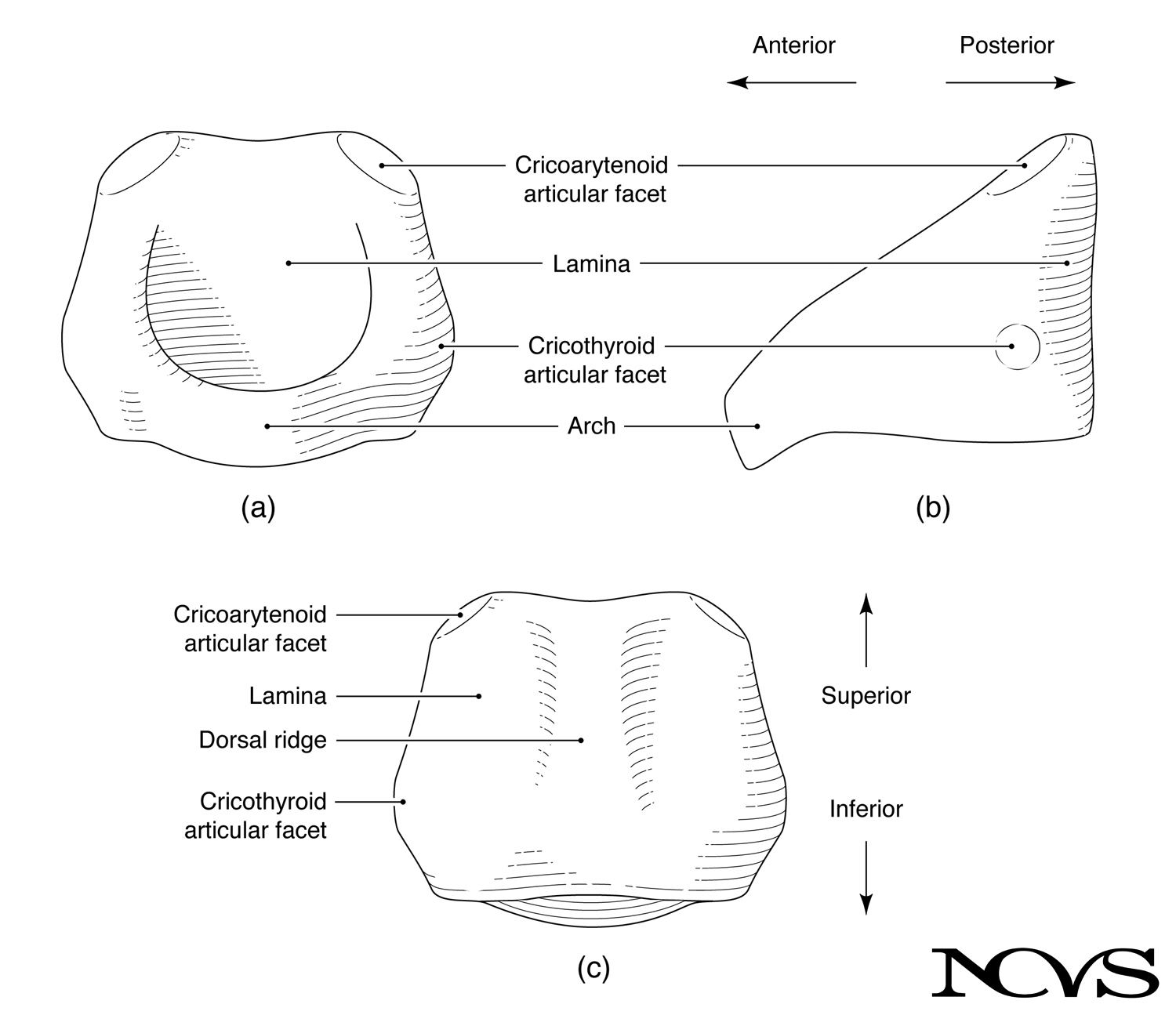 Figure 1.5: The cricoid cartilage shown in (a) anterior view, (b) lateral view, and (c) posterior view.
Structural Features
The cricoid may be thought of as the most superior tracheal ring, but it is different in shape and unique in its 360° solid construction. The cricoid resembles a signet ring: it is taller posteriorly (forming the lamina) and shorter anteriorly (forming the arch), as seen in Figure 1.5b.
This signet ring shape has important functional implications. The taller posterior portion provides a stable platform for the arytenoid cartilages, which sit atop the posterior cricoid lamina. The anterior arch can move upward toward the thyroid cartilage when rotation about the cricothyroid joint takes place.
Articular Surfaces
The cricoid cartilage has two types of smooth articular facets:
Cricothyroid Articular Facets: Small, smooth surfaces located laterally on each side, where the inferior cornua of the thyroid cartilage attach to form the cricothyroid joint (Figure 1.5a, b). This joint enables the rocking motion between thyroid and cricoid cartilages that is essential for vocal fold lengthening and pitch control.
Cricoarytenoid Articular Facets: Smooth surfaces on top of the posterior cricoid lamina, where the arytenoid cartilages attach (Figure 1.5c). These facets form the cricoarytenoid joints, which enable the complex rocking and gliding motions of the arytenoid cartilages necessary for vocal fold abduction and adduction.
From a posterior view, the cricoid appears as a hexagonal plate with a dorsal ridge at the midline (Figure 1.5c). This ridge provides attachment for muscles, particularly the longitudinal esophageal muscles.
Clinical Significance
Because the cricoid forms a complete ring, it is the narrowest point of the adult airway. In children, the cricoid region (subglottis) is the narrowest airway point, making this area critical in pediatric airway management. Edema or inflammation in the subglottic region can rapidly compromise breathing.
The cricoid’s structural integrity is essential—unlike the thyroid cartilage, which can be partially resected surgically, damage to the cricoid ring compromises airway stability.
The Arytenoid Cartilages
The arytenoid cartilages (paired) are pyramidal in shape and situated on top of the posterior portion of the cricoid lamina. These small but mechanically crucial cartilages are responsible for the dynamic positioning of the vocal folds.
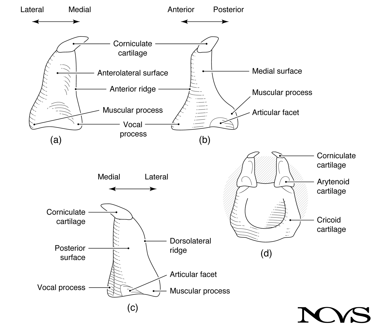 Figure 1.6: The arytenoid cartilage shown in (a) anterior view, (b) lateral view, (c) posterior view, and (d) composite anterior view with cricoid cartilage.
Structural Features
Each arytenoid has a pyramidal shape with three surfaces (anterolateral, medial, posterior), a base, and an apex. The base sits on the cricoid lamina at the cricoarytenoid articular facet. The apex points superiorly and slightly posteriorly, often capped by the small corniculate cartilage (cartilage of Santorini).
At the base of each arytenoid cartilage are two important projections:
Vocal Process: Projects anteriorly from the base (Figure 1.6a, b). This is the attachment point for the vocal ligament, making it the posterior anchor of the vocal fold. The position of the vocal process directly determines vocal fold length and tension.
Muscular Process: Projects laterally from the base (Figure 1.6a, b). This serves as the attachment point for the lateral cricoarytenoid and posterior cricoarytenoid muscles, which abduct and adduct the vocal folds.
The arytenoid surfaces also serve specific functions. The medial surface faces the opposite arytenoid across the midline. When the vocal folds are adducted, these medial surfaces approximate, helping to seal the glottis. The anterolateral surface is somewhat concave and provides attachment for the thyroarytenoid muscle (the body of the vocal fold).
Joint Mechanics
The cricoarytenoid joint is a highly mobile synovial joint with complex kinematics. The articular facet on the cricoid is curved, allowing a rocking motion (rotation and translation) of the arytenoid cartilage on top of the cricoid (Figure 1.6c, d).
This rocking motion enables the arytenoid cartilages to move in multiple directions:
- Medial-lateral: bringing the vocal processes together (adduction) or apart (abduction)
- Anterior-posterior: moving the vocal processes forward or backward
- Rotational: pivoting around the vertical axis
The exact degrees of freedom throughout the rocking movement have been debated, with some researchers emphasizing sliding motions and others emphasizing rotational components. Most current evidence suggests the cricoarytenoid joint combines both rotation and translation, enabling the complex three-dimensional positioning necessary for vocal fold control.
Figure 1.7: Vocal folds viewed with videofiberscopy showing (a) abduction and (b) adduction. The arytenoid cartilages are near the bottom of each image. Photos by Debra K. Klein, The University of Iowa Hospitals and Clinics.
Functional Significance
The arytenoid cartilages are arguably the most important cartilages for voice production. Their position determines:
- Glottal opening size: affecting airflow and resistance
- Vocal fold length: affecting fundamental frequency
- Vocal fold tension: affecting loudness and vocal quality
- Glottal closure pattern: affecting voice quality and efficiency
Paralysis or fixation of an arytenoid cartilage (often resulting from recurrent laryngeal nerve damage) severely impairs voice production and can compromise breathing.
The Epiglottis
The epiglottis is a leaf-shaped elastic cartilage that resembles the tongue of a shoe. Like the tongue of a shoe folds over the foot when laced, the epiglottis folds over the entryway to the larynx when tight closure of the airway is desired.
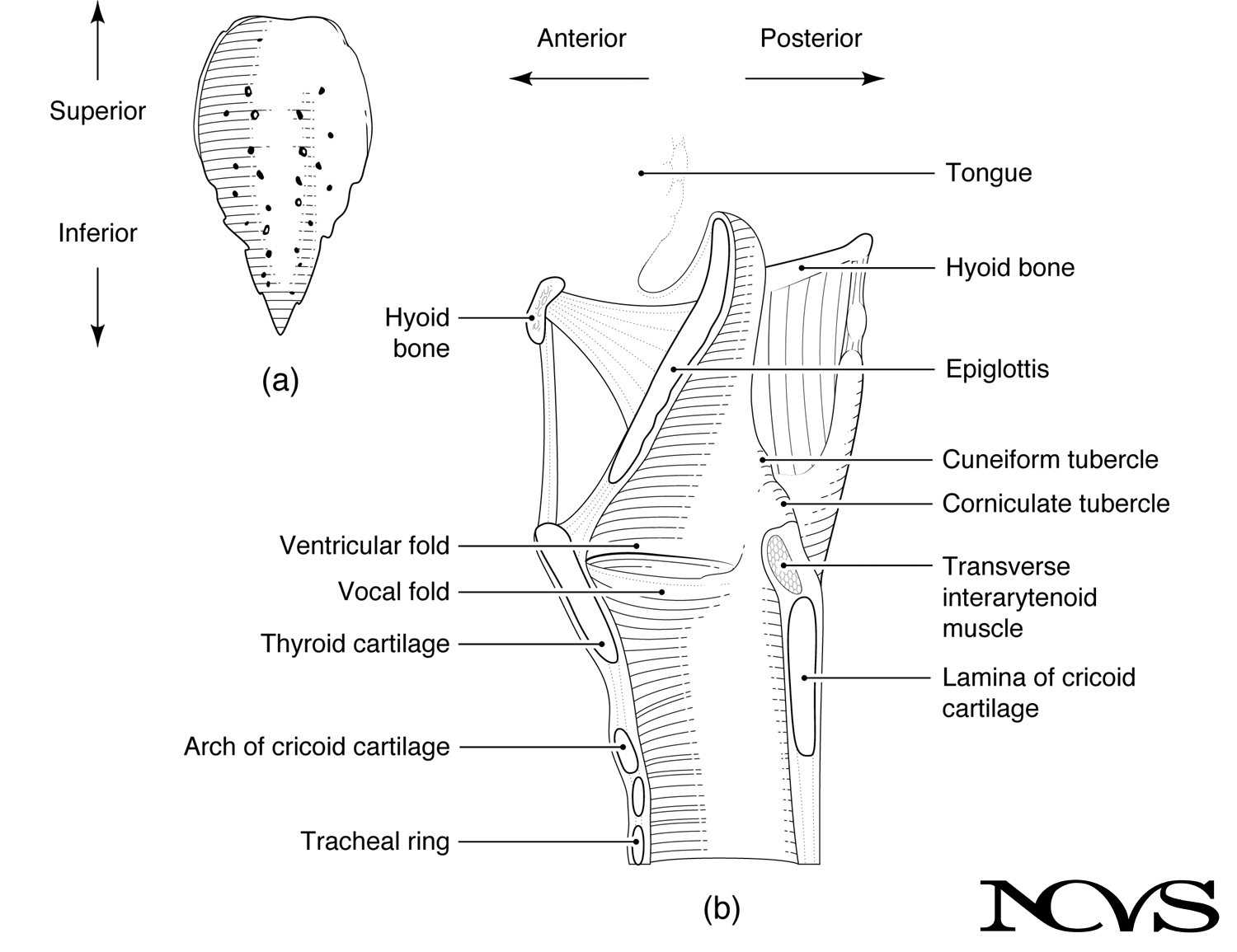 Figure 1.8: The epiglottis shown in (a) posterior view and (b) midsagittal section through the larynx, revealing attachments to the hyoid bone, tongue, and thyroid cartilage.
Structural Features and Attachments
The epiglottis is composed of elastic cartilage, which does not ossify with age and maintains flexibility throughout life. Its broad superior portion is free and projects upward behind the tongue. Its narrow inferior portion (the petiole or stem) attaches to the inner surface of the thyroid cartilage, just below the thyroid notch (Figure 1.8b).
The epiglottis has multiple attachment points:
Thyroid Cartilage: The petiole connects via the thyroepiglottic ligament to the internal midline of the thyroid cartilage, anchoring the epiglottis anteriorly.
Hyoid Bone: The anterior surface of the epiglottis connects to the posterior surface of the hyoid bone via the hyoepiglottic ligament (Figure 1.8b).
Tongue: The anterior surface also connects to the base of the tongue via the glossoepiglottic folds (median and lateral).
Functional Roles
Airway Protection: During swallowing, the larynx elevates and moves anteriorly while the epiglottis tilts posteriorly and inferiorly, creating a protective covering over the laryngeal opening. This movement redirects the food bolus into the lateral channels (pyriform sinuses) on either side of the larynx.
Acoustic Resonator: When the airway is open during phonation, the epiglottis and surrounding structures form the epiglottal chamber, a small acoustic space between the epiglottis and the posterior pharyngeal wall. This chamber can serve as an acoustic resonator, particularly in certain voice qualities and in some phonetic contexts (see Chapter 9).
Clinical Note
Inflammation of the epiglottis (epiglottitis) can be life-threatening, particularly in children, as a swollen epiglottis can obstruct the airway. This condition is now rare due to vaccination against Haemophilus influenzae type B, which was the primary cause.
Additional Small Cartilages
Two additional pairs of small cartilages are present in the larynx:
Corniculate Cartilages (Cartilages of Santorini): Small, conical elastic cartilages that sit atop the apex of each arytenoid cartilage (Figure 1.6a, d). They are often fused with the arytenoid apex and may help support the aryepiglottic folds.
Cuneiform Cartilages (Cartilages of Wrisberg): Small, rod-shaped elastic cartilages embedded in the aryepiglottic folds (the tissue folds connecting the arytenoids to the epiglottis). They provide structural support to these folds and are sometimes visible as small bumps on the lateral walls of the larynx during endoscopy (Figure 1.8b).
Summary
The laryngeal cartilages form an elegantly engineered framework that balances stability with mobility. The thyroid cartilage provides a protective shield and serves as an anchor for the vocal folds anteriorly. The cricoid cartilage forms a complete ring that maintains airway patency and provides the foundation for arytenoid movement. The arytenoid cartilages enable the fine motor control of vocal fold positioning essential for voice production. The epiglottis protects the airway during swallowing while potentially contributing to acoustic resonance during phonation.
Understanding these cartilaginous structures and their articulations is fundamental to comprehending how muscles act upon this framework to control breathing, phonation, and airway protection. The joints between these cartilages—particularly the cricothyroid and cricoarytenoid joints—are critical pivot points that enable the larynx to serve its multiple functions.
Key Takeaways
- ✅ The thyroid cartilage forms the anterior-lateral laryngeal wall and exhibits sexual dimorphism in angle and size
- ✅ The cricoid is the only complete ring cartilage, forming the foundation of the laryngeal framework
- ✅ The arytenoid cartilages control vocal fold position through complex three-dimensional movements at the cricoarytenoid joints
- ✅ The epiglottis protects the airway during swallowing and may serve as an acoustic resonator during phonation
- ✅ Cricoarytenoid and cricothyroid joints enable the mobility essential for pitch control and vocal fold positioning
- ✅ Corniculate and cuneiform cartilages provide structural support to the laryngeal folds and opening
Related Topics
Further Reading
- Judson, L. S., & Weaver, A. T. (1942). Voice science. New York: F. S. Crofts.
- Fink, B. R. (1975). The human larynx: A functional study. New York: Raven Press.
- Von Leden, H., & Moore, P. (1961). The mechanics of the cricoarytenoid joint. Archives of Otolaryngology, 73, 541-550.
- Romanes, G. J. (1964). Cunningham’s textbook of anatomy. Oxford University Press.