Vascularity and Innervation of the Larynx
The larynx requires a robust blood supply to sustain the metabolic demands of its muscles and to support tissue repair following the mechanical stresses of phonation. Similarly, precise neural control is essential for the coordinated muscle activity necessary for breathing, phonation, and airway protection. This topic provides a brief overview of the vascular and neural pathways that support laryngeal function.
Arterial Blood Supply
The larynx receives blood primarily from two arteries, both of which are branches of the carotid system. These vessels form an interconnected network (anastomosis) ensuring redundant blood supply to critical structures.
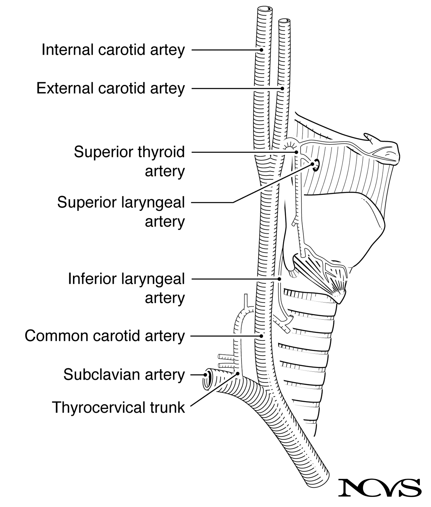 Figure 1.16: Major arteries supplying blood to the larynx, showing the superior and inferior laryngeal arteries branching from the thyroid arteries.
Superior Laryngeal Artery
The superior laryngeal artery arises from the superior thyroid artery, which itself branches from the external carotid artery. The superior laryngeal artery accompanies the internal branch of the superior laryngeal nerve, piercing the thyrohyoid membrane to enter the larynx.
Distribution:
- Supplies the supraglottic larynx (structures above the vocal folds)
- Provides blood to the epiglottis, aryepiglottic folds, and ventricular folds
- Anastomoses (connects) with branches from the inferior laryngeal artery
The superior laryngeal artery’s course through the thyrohyoid membrane makes it susceptible to injury during certain surgical procedures in this region.
Inferior Laryngeal Artery
The inferior laryngeal artery arises from the inferior thyroid artery, which branches from the thyrocervical trunk (itself arising from the subclavian artery). The inferior laryngeal artery ascends alongside the recurrent laryngeal nerve, entering the larynx from below.
Distribution:
- Supplies the subglottic larynx and the posterior and lateral aspects of the vocal folds
- Provides blood to the intrinsic laryngeal muscles
- Anastomoses with branches from the superior laryngeal artery
Transarterial Cascade and Microcirculation
Within the larynx, the superior and inferior laryngeal arteries connect via a transarterial cascade, forming an interconnected network. From this network, smaller branches lead to various laryngeal structures and muscles.
Within the vocal folds themselves, tiny blood vessels (capillaries) run primarily in the anterior-posterior direction, along the tissue fibers. This orientation has prompted interesting speculation about the relationship between vocal fold vibration and blood flow.
Potential Effects of Vibration on Blood Flow:
Does the repeated acceleration and deceleration of tissue during phonation affect blood flow? At what point can vibration or tissue compression cause a vessel to rupture? These are questions that remain incompletely answered.
One hypothesis is that high-amplitude, high-frequency vibration might temporarily reduce blood flow to the vocal folds, similar to how sustained muscle contraction can restrict blood flow to working muscles. If this occurs during prolonged or intense voice use, it could contribute to vocal fatigue. However, the vocal folds also benefit from rest periods during normal speech (between utterances), which may allow for blood flow recovery.
Vocal Fold Hemorrhage: Rupture of blood vessels within the vocal fold can occur from excessive mechanical stress, particularly during forceful phonation with improper technique. Vocal fold hemorrhage typically involves rupture of capillaries in the superficial lamina propria, causing acute voice loss and requiring strict voice rest for healing.
Venous Drainage
Venous blood from the larynx drains through veins that parallel the arterial supply:
Superior Laryngeal Vein: Drains to the superior thyroid vein, which empties into the internal jugular vein.
Inferior Laryngeal Vein: Drains to the inferior thyroid vein, which empties into the brachiocephalic vein (left side) or internal jugular vein (right side).
The venous system removes metabolic waste products and deoxygenated blood from laryngeal tissues. Venous congestion or vascular lesions (such as ectasias or varices) can sometimes be visualized during laryngoscopy and may contribute to voice problems.
Innervation of the Larynx
Laryngeal innervation is accomplished through two major branches of the vagus nerve (cranial nerve X): the superior laryngeal nerve and the recurrent laryngeal nerve. These nerves provide both motor innervation (controlling muscle contraction) and sensory innervation (providing feedback about touch, position, and chemical stimuli).
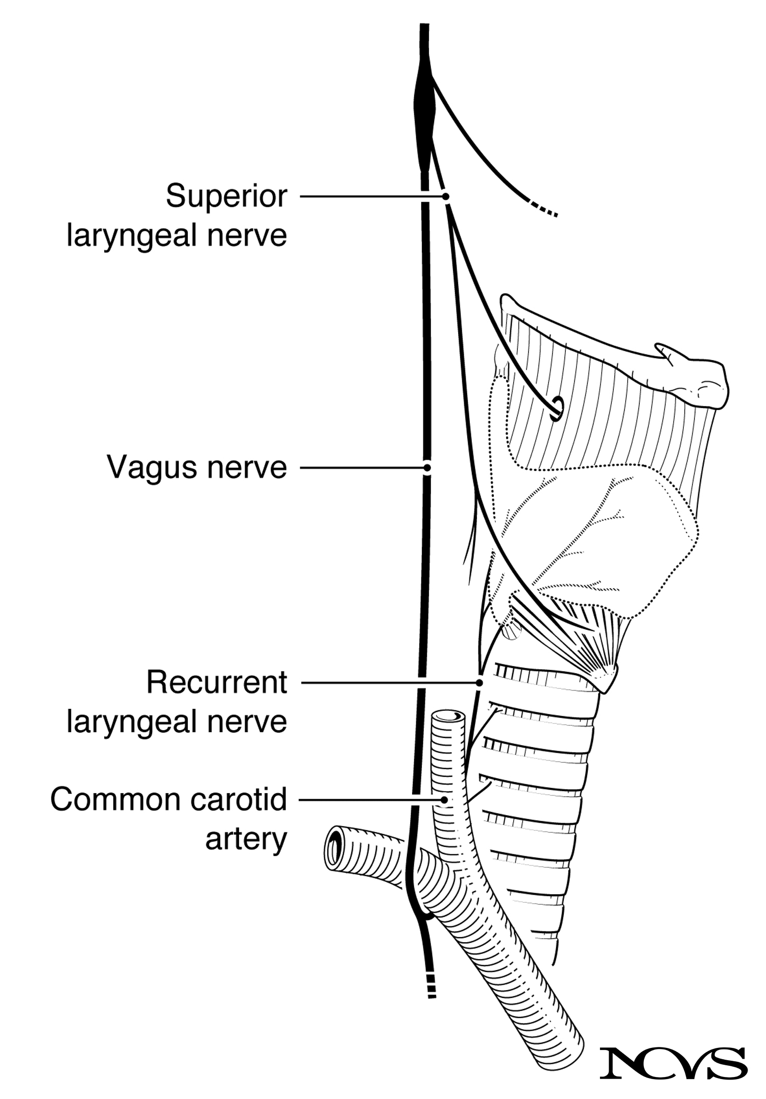 Figure 1.17: Innervation of the larynx, showing various branches splitting off the vagus nerve.
The Vagus Nerve
The vagus nerve (cranial nerve X) originates in the brainstem (medulla oblongata) and descends through the neck alongside the carotid artery. It is part of the parasympathetic nervous system and has extensive distributions to the heart, lungs, and digestive system, in addition to the larynx.
The vagus gives off two critical branches for laryngeal function:
- Superior laryngeal nerve (arises high in the neck)
- Recurrent laryngeal nerve (arises in the chest and “recurs” upward)
Superior Laryngeal Nerve
The superior laryngeal nerve branches from the vagus nerve high in the neck, near the level of the hyoid bone. It divides into two branches:
Internal Branch
The internal branch of the superior laryngeal nerve is primarily sensory. It pierces the thyrohyoid membrane (along with the superior laryngeal artery) to enter the larynx.
Sensory Distribution:
- Provides sensation to the supraglottic larynx (epiglottis, aryepiglottic folds, laryngeal surface above the vocal folds)
- Mediates the laryngeal adductor reflex: when irritating stimuli contact the supraglottic larynx, the vocal folds close reflexively to protect the airway
- Contributes to the cough reflex
Loss of sensation from internal branch damage can increase aspiration risk, as the protective reflexes may not trigger appropriately when foreign material enters the larynx.
External Branch
The external branch of the superior laryngeal nerve is primarily motor. It courses along the lateral surface of the inferior pharyngeal constrictor muscle and reaches the cricothyroid muscle.
Motor Function:
- Innervates the cricothyroid muscle exclusively
- Provides the neural control for vocal fold elongation and pitch elevation
The cricothyroid muscle is the only intrinsic laryngeal muscle innervated by the superior laryngeal nerve. This isolated innervation makes the function of the cricothyroid—raising pitch by vocal fold elongation—very selective and functionally independent from other laryngeal adjustments.
Superior Laryngeal Nerve Paralysis: Damage to the external branch of the superior laryngeal nerve results in paralysis of the cricothyroid muscle. Patients typically report:
- Difficulty reaching high pitches
- Vocal fatigue during prolonged speaking
- Decreased vocal range
- Subtle voice quality changes (often described as “hoarseness” or reduced projection)
The voice may sound relatively normal in casual conversation but deteriorates with increased vocal demands.
Recurrent Laryngeal Nerve
The recurrent laryngeal nerve has an unusual anatomical course. After the vagus nerve descends into the thorax (chest), the recurrent laryngeal nerve branches off and loops back upward to innervate the larynx.
Anatomical Course:
Left Side: The left recurrent laryngeal nerve loops under the aortic arch (the main artery leaving the heart) before ascending in the groove between the trachea and esophagus to reach the larynx.
Right Side: The right recurrent laryngeal nerve loops under the subclavian artery before ascending.
The nerve is called “recurrent” because it passes along (and then returns to) the larynx. After splitting from the vagus, it courses downward along the neck into the thorax and returns to enter the larynx from the rostral-caudal direction (from below, moving upward).
Motor Function
The recurrent laryngeal nerve provides motor innervation to all intrinsic laryngeal muscles except the cricothyroid:
- Thyroarytenoid (vocal fold body)
- Lateral cricoarytenoid (adductor)
- Posterior cricoarytenoid (abductor)
- Interarytenoid (adductor)
This shared innervation requires intricate neural control to coordinate the various opposing muscle actions necessary for phonation and breathing.
Sensory Function
The recurrent laryngeal nerve also provides sensory innervation to the larynx below the level of the vocal folds (subglottic larynx and trachea).
Branching and Muscle Specificity
As the recurrent laryngeal nerve ascends into the larynx, it branches to innervate different muscles. The exact branching pattern has been debated, particularly regarding the posterior cricoarytenoid and lateral cricoarytenoid muscles, which are agonist-antagonist pairs (one abducts, the other adducts the vocal folds).
The intricate branching must allow for independent control of these opposing muscles. Some researchers have proposed that different branches of the recurrent laryngeal nerve control different muscle groups, enabling selective activation. This topic remains an area of active research.
Recurrent Laryngeal Nerve Paralysis
Recurrent laryngeal nerve paralysis is one of the most common and significant laryngeal nerve injuries. It can result from:
- Surgical trauma (thyroid surgery, cardiac surgery, cervical spine surgery)
- Tumors compressing the nerve (lung cancer, thyroid cancer, mediastinal masses)
- Viral infections
- Idiopathic causes (unknown etiology)
Unilateral (One-Sided) Paralysis:
- The affected vocal fold assumes a paramedian or lateral position (partway between closed and fully abducted)
- Breathy voice due to incomplete glottal closure
- Reduced vocal loudness
- Possible aspiration (liquid entering the airway), particularly with thin liquids
- Some patients compensate well; others have severe voice impairment
Bilateral (Both Sides) Paralysis:
- Both vocal folds are immobilized, typically in a paramedian position
- Stridor (noisy breathing) due to narrowed airway
- Respiratory distress, potentially requiring emergency tracheostomy
- Voice may be relatively preserved (if vocal folds are close together) but at the cost of compromised breathing
- Treatment often involves surgically widening the airway (arytenoidectomy or cordotomy), which improves breathing but worsens voice
The recurrent laryngeal nerve’s long, circuitous path makes it vulnerable to injury at multiple points, which is why laryngeal nerve paralysis is a relatively common clinical problem.
Agonist-Antagonist Control and Neural Complexity
The recurrent laryngeal nerve must provide differential control to multiple pairs of agonist-antagonist muscles:
Abduction vs. Adduction:
- Posterior cricoarytenoid (abductor) vs. lateral cricoarytenoid and interarytenoid (adductors)
Shortening vs. Lengthening (in combination with superior laryngeal nerve):
- Thyroarytenoid (shortener/thickener) vs. cricothyroid (lengthener)
The nervous system controls these opposing forces through reciprocal innervation—when one muscle group is activated, the antagonist is typically inhibited (though not always completely). Fine motor control involves continuously adjusting the balance between agonist and antagonist activity.
This neural control is not merely “on/off” but involves graded changes in motor unit recruitment and firing rates, allowing for the subtle adjustments necessary for speech prosody, singing, and emotional expression through voice.
Summary
The larynx receives blood supply from the superior and inferior laryngeal arteries, which form an interconnected network ensuring adequate perfusion of all laryngeal structures. Tiny blood vessels within the vocal folds themselves run longitudinally, and there is ongoing interest in how vocal fold vibration might affect blood flow and contribute to vocal fatigue or vascular injury.
Innervation is accomplished through two branches of the vagus nerve. The superior laryngeal nerve provides sensory innervation to the supraglottic larynx (internal branch) and motor innervation to the cricothyroid muscle (external branch). The recurrent laryngeal nerve provides motor innervation to all other intrinsic muscles and sensory innervation to the subglottic larynx.
The isolated innervation of the cricothyroid by the superior laryngeal nerve makes pitch control functionally independent from other laryngeal adjustments. The recurrent laryngeal nerve’s innervation of multiple agonist-antagonist muscle pairs requires intricate neural control for coordinated laryngeal function.
Understanding vascular and neural anatomy is essential for comprehending voice physiology, interpreting clinical findings, and anticipating the consequences of surgical interventions or pathological processes affecting these structures.
Key Takeaways
- ✅ The superior laryngeal artery supplies the supraglottic larynx; the inferior laryngeal artery supplies the subglottic region and vocal folds
- ✅ Blood vessels within vocal folds run longitudinally, potentially affected by vibratory forces during phonation
- ✅ The superior laryngeal nerve has internal (sensory) and external (motor to cricothyroid) branches
- ✅ The recurrent laryngeal nerve innervates all intrinsic muscles except cricothyroid and provides subglottic sensation
- ✅ Recurrent laryngeal nerve paralysis causes vocal fold immobility, resulting in breathy voice (unilateral) or respiratory distress (bilateral)
- ✅ The long course of the recurrent laryngeal nerve makes it vulnerable to injury from surgery, tumors, or other pathology
Related Topics
- Intrinsic Muscles of the Larynx
- Extrinsic Muscles of the Larynx
- Laryngeal Cartilages
- Morphology of Vocal Fold Soft Tissue
Further Reading
- Cooper, M. (1971). Papillomata of the vocal folds: A review. Journal of Speech and Hearing Research, 36, 51-60.
- Fink, B. R. (1975). The human larynx: A functional study. New York: Raven Press.
- Romanes, G. J. (1964). Cunningham’s textbook of anatomy. Oxford University Press.
- Zemlin, W. R. (1964). Speech and hearing science: Anatomy and physiology. Champaign, IL: Stipes Publishing Co.