Extrinsic Muscles of the Larynx
The extrinsic laryngeal muscles connect the larynx and hyoid bone to surrounding structures in the head, neck, and thorax. Unlike the intrinsic muscles, which adjust vocal fold configuration, the extrinsic muscles control the position of the entire laryngeal complex within the neck. This positioning affects pharyngeal space, vocal tract length, and the mechanical advantage of intrinsic muscles—all factors that influence voice quality and resonance.
Functional Overview
Extrinsic laryngeal muscles serve multiple functions:
Laryngeal Height Control: Raising or lowering the larynx in the neck during voice production, changing vocal tract length and pharyngeal space configuration.
Swallowing Coordination: Elevating the larynx and closing the airway to protect against aspiration during swallowing.
Airway Opening: Depressing and stabilizing the larynx during deep inspiration to maximize airflow.
Postural Stabilization: Providing a stable platform for intrinsic muscle action by fixing the hyoid bone and larynx.
The extrinsic muscles are typically divided into two groups: infrahyoid muscles (below the hyoid, generally depressing the larynx) and suprahyoid muscles (above the hyoid, generally elevating the larynx).
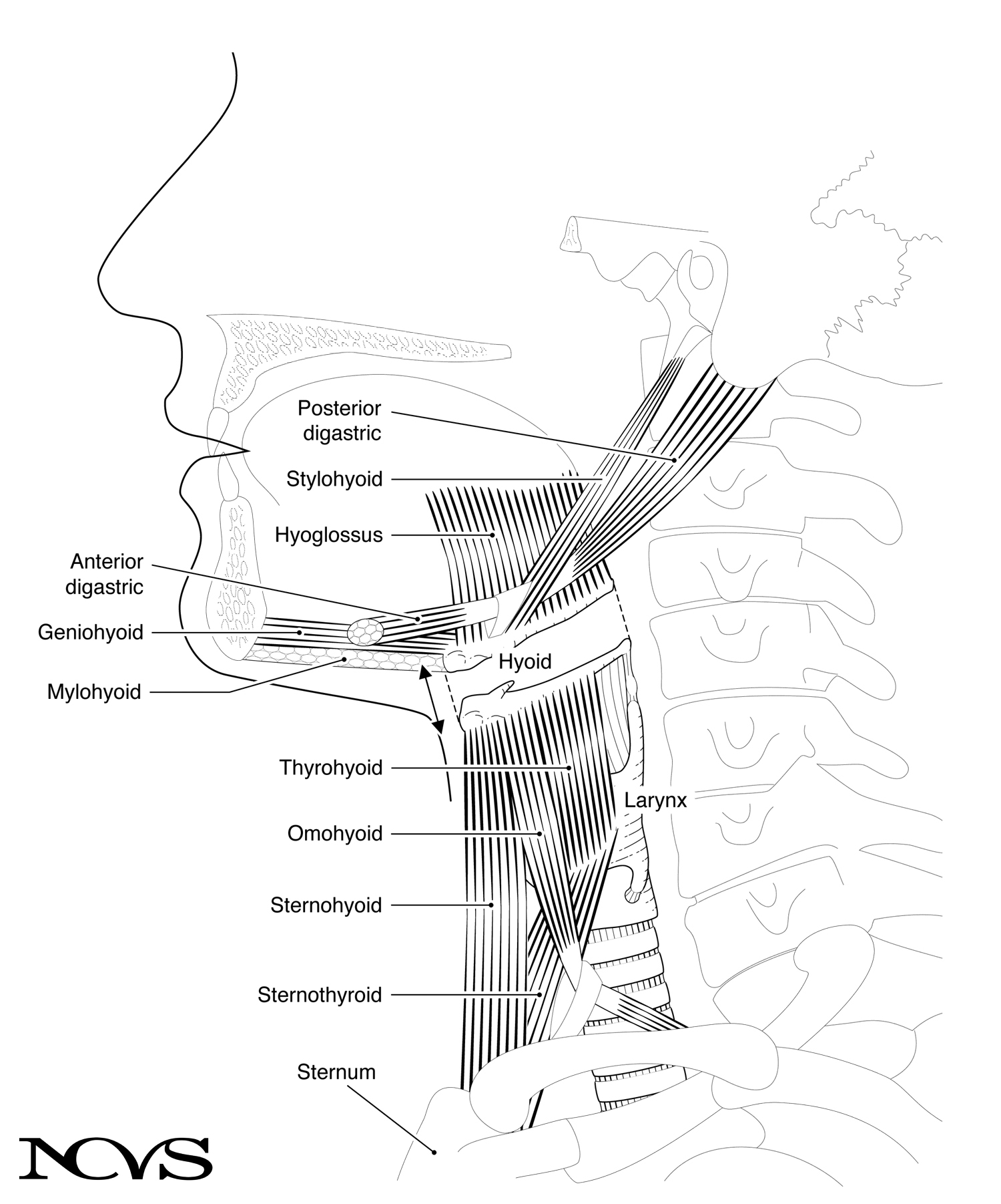 Figure 1.11: Extrinsic muscles of the larynx in relation to other head and neck structures.
Infrahyoid Muscles (Laryngeal Depressors)
The infrahyoid muscles are often called “strap muscles” because of their relatively flat, band-like appearance. They connect the larynx and hyoid to structures below (sternum, scapula), pulling the laryngeal complex downward.
Sternohyoid Muscle
The sternohyoid muscle (paired) runs vertically from the posterior surface of the sternum (breastbone) to the lower border of the hyoid bone (Figure 1.11).
Origin: Posterior surface of the manubrium of the sternum and medial end of the clavicle
Insertion: Inferior border of the hyoid body
Action: When contracted, shortens the distance between the sternum and hyoid bone. It is therefore a laryngeal depressor (indicated by a double-headed arrow in Figure 1.11), but lowering the larynx is indirect since it acts through connective tissue between the hyoid bone and the larynx to pull the larynx downward.
The sternohyoid can also stabilize the hyoid bone, preventing it from being pulled upward by suprahyoid muscles. This stabilization provides a fixed anchor for tongue movements during speech articulation.
Sternothyroid Muscle
The sternothyroid muscle (paired) originates at the posterior surface of the sternum, runs laterally upward, and attaches to the oblique line of the thyroid lamina (Figure 1.11).
Origin: Posterior surface of the manubrium of the sternum
Insertion: Oblique line on the thyroid lamina
Action: The sternothyroid muscle lowers (depresses) the larynx in a direct way. Unlike the sternohyoid, which acts indirectly through the hyoid, the sternothyroid attaches directly to the laryngeal framework. It is the most direct laryngeal depressor.
During singing or speaking, the sternothyroid can maintain a lowered laryngeal position, which is associated with increased pharyngeal space and a “darker” or “rounder” vocal timbre. Some voice training methods emphasize controlled sternothyroid engagement to achieve specific timbral goals.
Thyrohyoid Muscle
The thyrohyoid muscle (paired) runs vertically from the oblique line of the thyroid cartilage toward the hyoid bone (Figure 1.11). It essentially continues the line of the sternothyroid upward.
Origin: Oblique line of the thyroid lamina
Insertion: Inferior border of the hyoid body and greater cornu
Action: The thyrohyoid’s action depends on which attachment is fixed:
- If the hyoid is fixed (stabilized by suprahyoid muscles), contraction elevates the larynx
- If the larynx is fixed (stabilized by sternothyroid and other infrahyoid muscles), contraction depresses the hyoid
During swallowing, the thyrohyoid often works in coordination with suprahyoid muscles to draw the larynx upward toward the hyoid, shortening the distance between them. During singing at high pitches, some voice pedagogy traditions suggest that thyrohyoid activity helps maintain connection between larynx and hyoid, potentially improving efficiency of cricothyroid muscle action.
Omohyoid Muscle
The omohyoid muscle (paired) has an unusual two-bellied structure connected by an intermediate tendon. It originates from the superior margin of the scapula (shoulder blade) and inserts into the inferior border of the hyoid bone (Figure 1.11).
Origin: Superior margin of the scapula
Insertion: Inferior border and lateral hyoid body
Action: The omohyoid depresses and retracts the hyoid bone. Its action is somewhat oblique due to its lateral origin, so it not only pulls the hyoid downward but also slightly backward.
The functional significance of the omohyoid is less clear than other infrahyoid muscles. It may play a role in stabilizing hyoid position during certain head and neck movements, or in coordinating laryngeal position with shoulder girdle posture.
Suprahyoid Muscles (Laryngeal Elevators)
The suprahyoid muscles connect the hyoid bone to structures above: the jaw (mandible), skull base, and tongue. While they are not strictly “laryngeal muscles” (they don’t attach directly to laryngeal cartilages), they affect laryngeal function by moving the hyoid, which in turn influences laryngeal position through the thyrohyoid membrane and related connective tissues.
Digastric Muscle
The digastric muscle (paired) has two bellies connected by an intermediate tendon that loops through a fibrous sling attached to the hyoid bone.
Anterior Belly:
- Origin: Digastric fossa on the inner surface of the mandible (near the midline)
- Action: Can retract the mandible (pull jaw backward) or elevate the hyoid
Posterior Belly:
- Origin: Mastoid notch of the temporal bone (behind the ear)
- Action: Elevates and retracts the hyoid bone
Combined Action: The two bellies perform somewhat opposing actions on the mandible—the posterior belly can retract it, and the anterior belly can depress it (if the hyoid is fixed). When both contract with the jaw fixed, they elevate the hyoid bone and thus indirectly elevate the larynx.
Stylohyoid Muscle
The stylohyoid muscle (paired) courses from the styloid process of the temporal bone to the hyoid bone (Figure 1.11).
Origin: Styloid process of the temporal bone (a thin, pointed projection of bone at the skull base)
Insertion: Body and greater cornu of the hyoid bone
Action: The stylohyoid elevates and retracts the hyoid bone, pulling it upward and backward. This action draws the larynx toward the base of the skull during swallowing and can be engaged during certain voice qualities that involve a retracted tongue root.
Mylohyoid Muscle
The mylohyoid muscle (paired) forms a muscular floor beneath the tongue, stretching between the two sides of the mandible like a hammock (Figure 1.11).
Origin: Mylohyoid line on the inner surface of the mandible
Insertion: Midline raphe and anterior hyoid body
Action: The mylohyoid elevates the hyoid when the mandible is fixed, or depresses the mandible when the hyoid is fixed. During swallowing, the mylohyoid elevates the hyoid and floor of the mouth, helping to propel the food bolus backward.
The mylohyoid also plays a role in tongue support. Its relationship to laryngeal position is indirect but clinically significant—excessive tension in the mylohyoid (sometimes called “tongue root tension”) can contribute to a raised laryngeal position and constricted voice quality.
Geniohyoid Muscle
The geniohyoid muscle (paired) runs from the inside of the chin to the hyoid bone (Figure 1.11).
Origin: Inferior mental spine (genial tubercle) on the inner surface of the mandible at the midline
Insertion: Anterior surface of the hyoid body
Action: The geniohyoid pulls the hyoid forward and upward when the mandible is fixed, or assists in mandible depression when the hyoid is fixed. During swallowing, this muscle contributes to the anterior and superior movement of the hyoid.
Hyoglossus Muscle
The hyoglossus muscle (paired) connects the hyoid bone to the lateral tongue.
Origin: Greater cornu and body of the hyoid bone
Insertion: Lateral aspect of the tongue
Action: The hyoglossus depresses the tongue or, if the tongue is fixed, elevates the hyoid. This muscle plays a significant role in tongue shaping during speech and singing, and its activity can influence laryngeal position through the hyoid connection.
Coordinated Extrinsic Muscle Function
Extrinsic muscles rarely work in isolation. Their coordinated activity creates the complex patterns of laryngeal movement necessary for speech, singing, and swallowing.
During Normal Speech
Laryngeal height varies continuously during connected speech, typically with small adjustments (a few millimeters) that correlate with pitch and phonetic context. The extrinsic muscles maintain a relatively neutral laryngeal position while making subtle adjustments:
- Infrahyoid muscles: Provide tonic baseline activity, preventing excessive elevation
- Suprahyoid muscles: Make rapid adjustments for pitch variation and articulatory demands
- Balance: Neither elevation nor depression predominates; larynx stays in mid-range
During Swallowing
Swallowing requires dramatic laryngeal elevation to close the airway and open the esophagus:
- Suprahyoid muscles contract strongly, elevating the hyoid bone
- Thyrohyoid contracts, pulling the larynx up toward the hyoid
- Infrahyoid muscles relax, allowing upward movement
- Epiglottis tilts posteriorly, covering the laryngeal inlet
- Upper esophageal sphincter opens, allowing food to enter
This coordinated sequence moves the larynx upward and forward by several centimeters, typically occurring in less than one second during a normal swallow.
During Singing
Voice pedagogues have long recognized that laryngeal position affects vocal quality and efficiency:
Elevated Larynx:
- Shortens the pharyngeal resonator
- Creates a “brighter,” more “forward” sound
- Associated with tension in some contexts, but can be functional in certain styles
- Common in untrained singers at high pitches
Lowered Larynx:
- Lengthens the pharyngeal resonator
- Creates a “darker,” “rounder,” or “covered” sound
- Often cultivated in classical voice training
- Requires controlled activity of infrahyoid muscles
Neutral Larynx:
- Generally preferred for speech-level singing and contemporary commercial music
- Allows natural adjustment to pitch without postural extremes
- Minimizes unnecessary muscular effort
The biomechanics of how laryngeal height affects voice production will be explored further in later chapters on resonance and vocal registration.
Clinical Significance
Dysfunction of extrinsic laryngeal muscles can significantly impact voice and swallowing:
Muscle Tension Dysphonia: Excessive extrinsic muscle tension (particularly suprahyoid muscles) can elevate the larynx and create a constricted, effortful voice quality. Treatment often involves manual therapy and voice exercises to reduce tension.
Paradoxical Vocal Fold Motion (PVFM): Sometimes associated with excessive laryngeal elevation during breathing, potentially involving suprahyoid muscle hyperactivity.
Dysphagia (swallowing difficulty): Weakness of suprahyoid muscles can reduce laryngeal elevation during swallowing, increasing aspiration risk. Therapy may target strengthening these muscles.
Presbyphagia (age-related swallowing changes): Muscle atrophy affects both intrinsic and extrinsic muscles, often reducing laryngeal elevation during swallowing.
Summary
The extrinsic laryngeal muscles form a complex suspension system that controls laryngeal position within the neck. Infrahyoid muscles (sternohyoid, sternothyroid, thyrohyoid, omohyoid) generally depress the larynx and stabilize the hyoid, while suprahyoid muscles (digastric, stylohyoid, mylohyoid, geniohyoid, hyoglossus) elevate the hyoid and indirectly raise the larynx.
The coordination between these muscle groups enables the larynx to be positioned optimally for breathing, swallowing, and voice production. During voice production, laryngeal height affects vocal tract length and pharyngeal space, influencing resonance and voice quality. During swallowing, dramatic laryngeal elevation protects the airway. Understanding extrinsic muscle function is essential for addressing voice disorders involving excessive laryngeal tension or inappropriate laryngeal positioning.
Key Takeaways
- ✅ Infrahyoid muscles (sternohyoid, sternothyroid, thyrohyoid, omohyoid) depress and stabilize the larynx
- ✅ Suprahyoid muscles (digastric, stylohyoid, mylohyoid, geniohyoid, hyoglossus) elevate the hyoid and indirectly raise the larynx
- ✅ The sternothyroid directly depresses the larynx; other infrahyoid muscles act through the hyoid
- ✅ Laryngeal height affects vocal tract length and pharyngeal space configuration, influencing voice quality
- ✅ Swallowing requires coordinated contraction of suprahyoid muscles and thyrohyoid to elevate the larynx
- ✅ Excessive extrinsic muscle tension can contribute to voice disorders and inefficient vocal production
Related Topics
Further Reading
- Palmer, J. M. (1984). Anatomy for speech and hearing (3rd ed.). New York: Harper & Row.
- Kahane, J., & Folkins, J. (1984). Atlas of speech and hearing anatomy. Columbus, OH: Charles E. Merrill Publishing.
- Zemlin, W. R. (1964). Speech and hearing science: Anatomy and physiology. Champaign, IL: Stipes Publishing Co.