Intrinsic Muscles of the Larynx
The intrinsic muscles of the larynx interconnect the laryngeal cartilages, with both origin and insertion on laryngeal structures. These muscles are the primary effectors of vocal fold control, directly determining glottal configuration, vocal fold length, and vocal fold tension. Understanding their anatomy and actions is fundamental to comprehending the biomechanics of phonation.
The Thyroarytenoid Muscle
The thyroarytenoid muscle (paired) courses from the thyroid cartilage (below the thyroid notch) to the arytenoid cartilage. It makes up the bulk of the vocal fold and is the largest intrinsic muscle by volume.
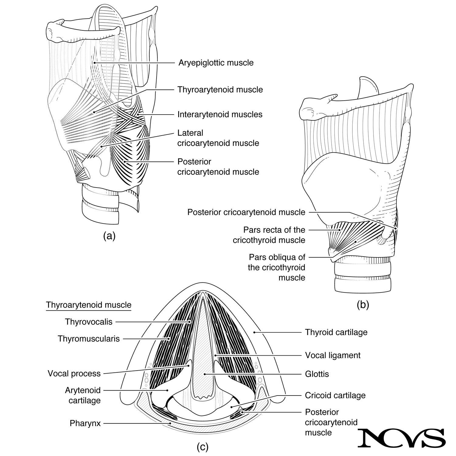 Figure 1.10: Intrinsic muscles shown in (a) posterior-lateral view and (b) anterior-lateral view, with (c) superior view revealing the thyroarytenoid muscle within the vocal folds.
Structural Organization
The thyroarytenoid muscle is sometimes divided into two functional bundles:
Thyrovocalis (or simply vocalis): The medial portion, consisting of the most medial fibers running along the length of the vocal fold. These fibers insert into the vocal ligament and lateral edge of the vocal fold. The vocalis may function in fine-tuning tension in localized regions of the vocal fold.
Thyromuscularis (or muscularis): The lateral portion, forming the bulk of the thyroarytenoid. These fibers run from the thyroid cartilage to the muscular process and body of the arytenoid cartilage.
The functional distinction between vocalis and muscularis is somewhat hypothetical but has biomechanical support. The vocalis may be used for fine-tuning the medial-most fibers during sustained phonation, while the muscularis may be used for quick shortening of the vocal folds during pitch changes or onset of phonation.
Actions
When the thyroarytenoid contracts:
- Draws the arytenoid cartilage forward, shortening the vocal folds
- Thickens the vocal folds by increasing their anterior-posterior dimension
- Increases the mass per unit length of the vibrating portion
- May stiffen the vocal fold body while allowing the cover to remain relatively lax
The muscle also stiffens in the process of contraction, contributing to control of fundamental frequency and vocal intensity. The thyroarytenoid works antagonistically with the cricothyroid muscle—while cricothyroid lengthens the vocal folds, thyroarytenoid shortens them.
Clinical Significance
The thyroarytenoid is the muscle most affected by aging-related atrophy (presbylaryngis), leading to vocal fold bowing and incomplete glottal closure. It is also a common target for botulinum toxin injection in the treatment of adductor spasmodic dysphonia, where weakening the muscle reduces excessive vocal fold compression.
The Cricothyroid Muscle
The cricothyroid muscle (paired) consists of two parts, both originating from the anterior arch of the cricoid cartilage:
Pars Recta (straight part): The more vertical portion courses upward to attach to the lower border of the thyroid lamina (Figure 1.10b).
Pars Obliqua (oblique part): The more horizontal portion runs upward and backward to attach to the inferior cornu of the thyroid cartilage (Figure 1.10b).
Actions and Biomechanics
The cricothyroid muscle is the primary pitch-control muscle. When it contracts, it produces a rocking motion at the cricothyroid joint:
- Elevates the anterior cricoid arch and/or depresses the thyroid lamina
- Increases the distance between the thyroid cartilage and the arytenoid cartilages
- Lengthens the vocal folds by pulling the thyroid cartilage forward and/or tilting it downward
- Increases longitudinal tension in the vocal fold tissues
- Elevates fundamental frequency (pitch)
The biomechanics will be explored in detail in Chapter 8, but the essential principle is that vocal fold elongation increases tension and thus increases the rate of vibration. The cricothyroid can produce vocal fold lengthening of approximately 3-6 mm in adults, resulting in pitch increases of up to one octave or more.
Functional Independence
The cricothyroid is the only intrinsic muscle innervated by the superior laryngeal nerve (external branch), rather than the recurrent laryngeal nerve. This isolated innervation makes pitch elevation functionally independent from glottal opening/closing and medial compression. A singer with recurrent laryngeal nerve paralysis can still adjust pitch (though voice quality will be severely affected by incomplete glottal closure).
Agonist-Antagonist Relationship
The cricothyroid and thyroarytenoid function as agonist-antagonist pairs for vocal fold length control:
- Cricothyroid: Lengthens and thins vocal folds
- Thyroarytenoid: Shortens and thickens vocal folds
The balance between these two muscles is crucial for pitch control and register transitions (chest voice vs. head voice). Skilled voice users learn to coordinate these muscles to maintain consistent voice quality across their pitch range.
The Lateral Cricoarytenoid Muscle
The lateral cricoarytenoid muscle (paired) originates from the superior borders of the lateral cricoid arch and inserts into the muscular process of the corresponding arytenoid cartilage (Figure 1.10a, c).
Actions
The lateral cricoarytenoid functions as an adductor of the vocal folds:
- Pulls the muscular process forward and medially
- Rotates the arytenoid cartilage, bringing the vocal processes together
- Closes the anterior glottis by approximating the vocal folds
The lateral cricoarytenoid brings together the vocal processes through a combination of rotation and forward rocking of the arytenoids on the cricoarytenoid joint. This action is essential for vocal fold closure during phonation.
Role in Glottal Closure
In combination with the interarytenoid muscle, the lateral cricoarytenoid achieves complete glottal closure. The lateral cricoarytenoid is particularly important for closing the anterior commissure—the junction where the left and right vocal folds meet anteriorly.
The Posterior Cricoarytenoid Muscle
The posterior cricoarytenoid muscle (paired) has its origin on the posterior surface of the cricoid cartilage. It courses upward and laterally to insert into the muscular process of the arytenoid cartilage (Figure 1.10a, c).
Actions
The posterior cricoarytenoid is the primary abductor of the vocal folds:
- Pulls the muscular process backward and laterally
- Rotates the arytenoid cartilage, moving the vocal processes apart
- Opens the glottis by separating the vocal folds
This muscle rotates and rocks the arytenoids backward, moving the vocal processes away from the midline. This action nearly opposes that of the lateral cricoarytenoid.
Critical Role in Breathing
The posterior cricoarytenoid is the only intrinsic muscle that opens the glottis. All other intrinsic muscles either close the glottis or have no effect on its width. This makes posterior cricoarytenoid function critical for breathing:
Bilateral posterior cricoarytenoid paralysis is a medical emergency, as the vocal folds cannot abduct for breathing. Patients may require immediate tracheostomy to establish an airway.
During normal breathing, the posterior cricoarytenoids abduct the vocal folds during inspiration, then allow partial adduction during expiration (under the influence of other muscles). During speech, these muscles must precisely control the degree of glottal opening for voicing versus voiceless sounds.
Agonist-Antagonist Relationship
The posterior cricoarytenoid and lateral cricoarytenoid are classic agonist-antagonist pairs:
- Posterior cricoarytenoid: Abducts (opens) the glottis
- Lateral cricoarytenoid: Adducts (closes) the glottis
The balance between these muscles determines glottal width and thus airflow resistance. For voice production, the glottis must be sufficiently closed to sustain oscillation, but not so tightly compressed that voice becomes strained or breaks.
The Interarytenoid Muscle
The interarytenoid muscle connects the two arytenoid cartilages posteriorly and is also considered to have two parts:
Transverse Part (unpaired): The transverse interarytenoid covers the entire posterior surface of the arytenoids. It originates on the lateral border of one arytenoid cartilage, runs horizontally across, and attaches to the lateral border of the other arytenoid (Figure 1.10a, c).
Oblique Part (paired): Each oblique interarytenoid originates from the muscular process of one arytenoid cartilage, courses diagonally upward, and inserts into the apex of the opposite arytenoid cartilage (Figure 1.10a). The oblique fibers cross each other like an “X” when viewed from behind.
Some fibers of the oblique interarytenoid continue beyond the arytenoid apex into the aryepiglottic fold, where they are sometimes called the aryepiglottic muscle. These fibers can draw the epiglottis posteriorly during swallowing.
Actions
The interarytenoid muscle serves as an adductor of the vocal folds:
- Draws the arytenoid cartilages together medially
- Closes the posterior glottis (the cartilaginous portion between the arytenoids)
- May assist in compressing the vocal folds along their entire length
While the lateral cricoarytenoid brings together the anterior glottis (membranous vocal folds), the interarytenoid muscle specifically seals off the posterior glottis. Complete glottal closure requires coordination of both muscle groups.
Role in the Anterior Commissure
Together with the lateral cricoarytenoid muscles, the interarytenoid ensures that the vocal folds meet along their entire length, including at the anterior commissure (where the vocal folds join in front). This complete closure is essential for:
- Efficient voice production (minimizing air leakage)
- Coughing and throat clearing (building subglottal pressure)
- Bearing down and lifting (Valsalva maneuver)
Incomplete glottal closure—whether from muscle weakness, scar tissue, or neurological dysfunction—results in breathy voice quality and reduced vocal efficiency.
Coordinated Muscle Actions
Voice production requires precisely coordinated activity across all intrinsic muscles. Consider the muscle activation patterns for different vocal tasks:
Quiet Breathing:
- Posterior cricoarytenoid: Active during inspiration (abducts vocal folds)
- Other intrinsic muscles: Minimal activity
Phonation (typical speech):
- Lateral cricoarytenoid and interarytenoid: Moderate activity (adduct vocal folds)
- Thyroarytenoid: Moderate activity (control stiffness and medial compression)
- Cricothyroid: Variable activity depending on pitch
- Posterior cricoarytenoid: Low tonic activity (balances adductory forces)
High-Pitched Phonation:
- Cricothyroid: High activity (lengthens vocal folds)
- Lateral cricoarytenoid and interarytenoid: High activity (maintains closure against increased tension)
- Thyroarytenoid: Reduced activity (allows lengthening but maintains some body stiffness)
Forced Glottal Closure (cough, Valsalva):
- Lateral cricoarytenoid, interarytenoid, and thyroarytenoid: Maximal activity
- Creates tight seal for pressure buildup
Summary
The intrinsic laryngeal muscles form an elegant system for controlling vocal fold configuration. The thyroarytenoid and cricothyroid work as antagonists to control vocal fold length and thickness. The lateral cricoarytenoid and posterior cricoarytenoid work as antagonists to control glottal width. The interarytenoid muscle seals the posterior glottis, working synergistically with the lateral cricoarytenoid to achieve complete glottal closure.
These muscles do not function in isolation but work in coordinated patterns determined by the task demands. The nervous system continuously adjusts the balance of agonist-antagonist activity to produce the subtle modulations necessary for speech prosody, singing, and other vocal behaviors. Understanding these individual muscles and their interactions provides the foundation for comprehending the biomechanics of phonation explored in later chapters.
Key Takeaways
- ✅ The thyroarytenoid muscle forms the body of the vocal fold and controls shortening and thickening
- ✅ The cricothyroid is the primary pitch-control muscle, lengthening the vocal folds through cricothyroid joint rotation
- ✅ The lateral cricoarytenoid adducts the vocal folds by pulling the muscular processes forward
- ✅ The posterior cricoarytenoid is the only glottal abductor, making it critical for breathing
- ✅ The interarytenoid muscle closes the posterior glottis and works synergistically with lateral cricoarytenoid for complete closure
- ✅ Cricothyroid and thyroarytenoid function as antagonists for length control; posterior and lateral cricoarytenoids function as antagonists for width control
Related Topics
Further Reading
- Hirano, M., & Kakita, Y. (1985). Cover-body theory of vocal cord vibration. In R. G. Daniloff (Ed.), Speech science (pp. 1-46). San Diego: College Hill Press.
- Von Leden, H., & Moore, P. (1961). The mechanics of the cricoarytenoid joint. Archives of Otolaryngology, 73, 541-550.
- Palmer, J. M. (1984). Anatomy for speech and hearing (3rd ed.). New York: Harper & Row.