Thoracic, Pleural, and Abdominal Pressure
Understanding pressure distribution throughout the torso is essential for comprehending respiratory function. While Pascal’s law provides a useful framework, biological tissues introduce complexities that require careful analysis.
Anatomical Organization
The Thorax
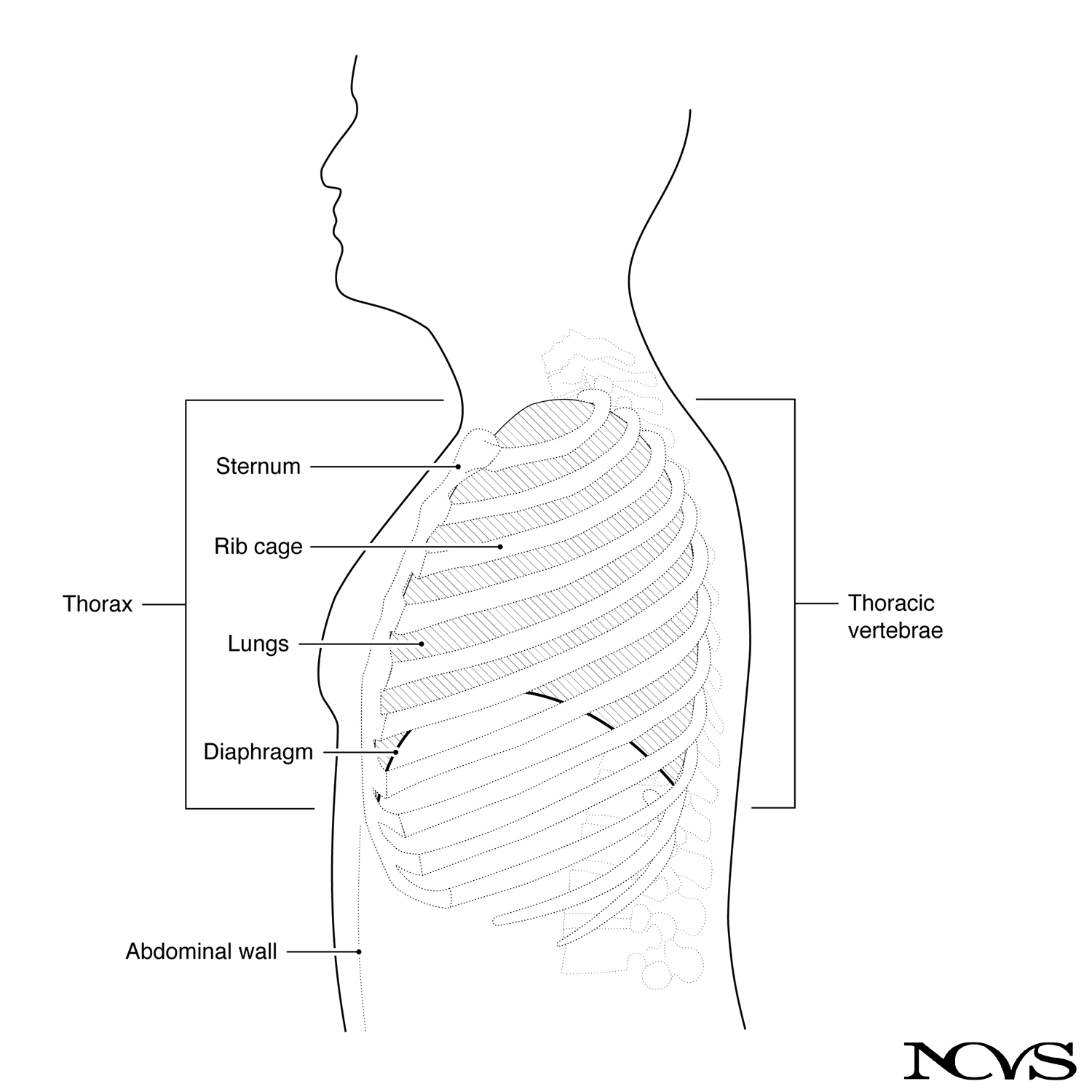 Figure 3.2: Diagram of the thorax and the abdomen. The thorax contains the rib cage, which houses the heart and the lungs.
The thorax is the region between the neck and abdomen. It contains the rib cage, which houses the lungs and heart. The thorax consists of bones, muscles, and connective tissue forming an outer shell for the torso above the midsection.
Major structural components include:
- Thoracic vertebrae (posterior)
- Sternum or breastbone (anterior)
- Ribs connecting vertebrae and sternum
- Ligaments and membranes providing additional support
The Diaphragm
The diaphragm is a large, dome-shaped structure of muscle and tendon located in the lower thorax, beneath the lungs. It divides the thorax from the viscera (the midsection containing stomach and intestines). The diaphragm plays a key role in breathing by changing thoracic volume.
The Pleural System
The lungs are doubly sealed within the rib cage, like a balloon within a slightly larger balloon:
- Visceral pleura: Inner membrane forming the outer walls of the lungs
- Parietal pleura: Outer membrane forming the inner wall of the rib cage
- Pleural space: The region between membranes, filled with liquid
This liquid-filled space allows the inner and outer membranes to slide relative to each other, like a thin layer of water between two balloons. This arrangement is crucial for smooth respiratory movements.
Pressure Transmission in the Torso
The Two-Fluid Model
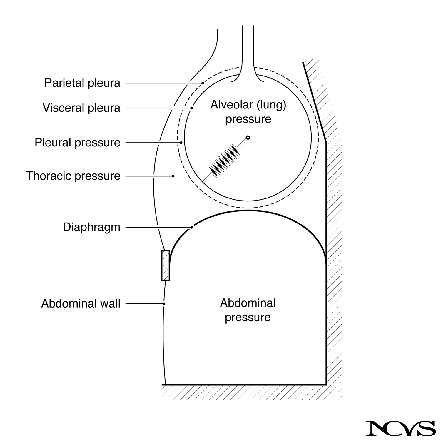 Figure 3.3: Highly schematized version showing how pressure is transmitted among abdominal, thoracic, and alveolar regions.
Assuming most biological tissues are liquid-like for pressure transmission, we can conceptually divide the torso into two major fluid spaces:
- Airspace: The continuous region within the alveoli
- Liquid space: All bones, membranes, ligaments, and other fibrous structures
This is an “impure” liquid in that Pascal’s law doesn’t apply directly. Only with modifications can the law describe pressure transmission through various parts of the torso.
Ideal Pressure Transmission
In the absence of gravity and elastic elements, pressure applied to the liquid space (by pushing against the torso or through muscle contraction) would transmit rapidly and uniformly throughout. This pressure would further transmit uniformly to the airspace, appearing as alveolar (lung) pressure.
Thus, one could theoretically manipulate lung pressure by pushing against either the thorax or the abdomen.
Factors Modifying Pressure Transmission
Gravitational Effects
Gravity acts on body tissues, making pressures in lower torso regions greater than upper regions in the upright position. The weight of body tissues contributes to increased pressure at the bottom, analogous to water pressure at the floor of a lake being determined by the column of water above.
As a result, abdominal pressures are generally greater than thoracic pressures in the upright position.
Elastic Recoil
Elastic structures—the visceral pleura, alveolar walls, and surface tension of liquid covering the alveoli—resist deformation. Think of the lungs as containing coiled springs like those in a mattress.
Any pressure applied to the mattress surface is resisted by the springs. If the mattress is sealed so air cannot escape, internal air pressure increases only to the extent that volume decreases. If coils strongly resist deformation, little pressure transmits to the inside.
Lung pressure is therefore determined by:
Lung pressure = Pleural pressure + Elastic recoil pressureWhen the “coils” (lung tissue) are extended, elastic recoil pressure adds to pleural pressure. When compressed, elastic recoil pressure subtracts from pleural pressure. Together, these pressures deform the lungs and change their volume.
Rib Cage Elasticity
The rib cage’s elasticity also affects pressure transmission. Pushing against the chest wall doesn’t transmit the same pressure to the lungs as pushing against the abdomen because of the rib cage’s greater stiffness compared to the abdominal wall.
Therefore, outer thoracic pressure differs from pleural pressure, even though a pure liquid model would predict them to be equal.
Practical Implications
Understanding these pressure relationships is crucial for:
- Breath support techniques: Manipulating thoracic versus abdominal effort
- Postural effects: How body position influences pressure distribution
- Muscle coordination: Balancing elastic forces with active muscle forces
- Clinical assessment: Interpreting pressure measurements in voice evaluation
The distinction between different pressures in thoracic and abdominal regions becomes essential in discussions of breathing mechanics and breath support strategies.
Summary
The torso contains multiple pressure regions that interact in complex ways. While Pascal’s law provides a foundation, gravitational and elastic effects create important deviations. Lung pressure results from the combined effects of pleural pressure and elastic recoil. Understanding these relationships is fundamental to analyzing respiratory function during phonation.
Key Takeaways
- ✅ The thorax contains the rib cage and is bounded below by the diaphragm
- ✅ The pleural system creates a double-sealed structure with liquid-filled space
- ✅ Gravity creates pressure gradients, with higher pressures in lower body regions
- ✅ Elastic recoil of lung tissue and rib cage modifies pressure transmission
Related Topics
Further Reading
- Hixon, T. J. (1973). Respiratory function in speech. In F. Minifie, T. Hixon, & F. Williams (Eds.), Normal aspects of speech, hearing, and language (pp. 73-125). Englewood Cliffs, NJ: Prentice Hall.
- Comroe, T. J., Jr. (1965). Physiology of respiration. Chicago: Year Book Medical Publishers.
- Dickson, D. R., & Maue-Dickson, W. (1982). Anatomical and physiological bases of speech. Boston: Little, Brown.