Normal Modes of Vibration in Vocal Fold Tissues
Increasing flexibility in tissue movement offers greater potential for achieving self-sustained oscillation. This flexibility can be expressed through the degrees of freedom of the tissue—the number of independent ways the system can move. Understanding normal modes of vibration reveals how the vocal folds select specific movement patterns optimized for energy transfer.
Degrees of Freedom
A degree of freedom represents one independent way a system can move. For the vocal folds:
Single-Mass Model:
- Each vocal fold has 1 degree of freedom for lateral (medial-lateral) motion
- Total: 2 degrees of freedom for both folds
- Cannot represent nonuniform movement
Two-Mass Model:
- Each vocal fold has 2 degrees of freedom (upper and lower masses)
- Total: 4 degrees of freedom
- Can represent vertical phase differences
Extended Models:
- Adding vertical displacement: 2 degrees of freedom per mass
- Adding anterior-posterior variation: Additional degrees per segment
- Real soft tissue: Infinite degrees of freedom (continuous deformable medium)
As more degrees of freedom are added, the system can exhibit more complex and realistic vibration patterns. However, most vibratory energy typically concentrates in a few dominant modes.
Rotational Degrees
Rigid bodies can also rotate about three perpendicular axes, adding up to 6 total degrees of freedom (3 translational + 3 rotational). For vocal fold vibration:
- Translational degrees (horizontal and vertical) are primary
- Rotational degrees (spinning or tumbling) are less important
- Focus remains on medial-lateral and inferior-superior translation
The Ribbon Model
The vocal fold cover can be conceptualized as a ribbon fixed at both ends (arytenoid and thyroid cartilages) but free to bend and flex in the middle. This model emphasizes:
- Longitudinal (anterior-posterior) variation in amplitude
- Wavelike motion along the length
- Continuous rather than discrete elements
- Similarity to vibrating strings or membranes
Normal modes describe the characteristic spatial patterns that such a ribbon can adopt during vibration.
Longitudinal Modes (Anterior-Posterior)
Consider modes varying along the length of the vocal fold from anterior to posterior attachment points.
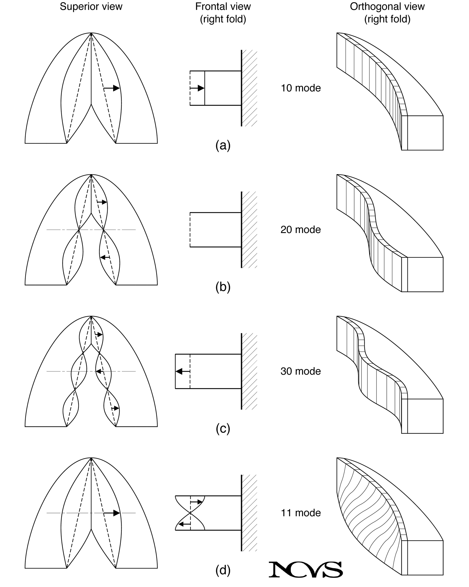 Figure 4.9: Normal modes showing (a) the 10 mode with maximum amplitude at midpoint, (b) the 20 mode with node at center, (c) the 30 mode with two nodes, and (d) the 11 mode with vertical phase difference.
Mode Notation
Modes are designated by integers mn where:
- m = number of half-wavelengths in the longitudinal direction
- n = number of half-wavelengths in the vertical direction
The 10 Mode
The fundamental longitudinal mode (m = 1, n = 0):
Characteristics:
- Amplitude maximum at midpoint of fold
- Amplitude decreases toward endpoints
- No vertical phase variation (uniform top to bottom)
- One half-wavelength spans the fold length
Physical Interpretation:
- Entire fold moves laterally in phase
- Similar to fundamental mode of a vibrating string
- Most common mode in simple mass-spring models
The 20 Mode
The second longitudinal mode (m = 2, n = 0):
Characteristics:
- Center of fold remains stationary (node)
- Anterior and posterior portions move in opposite directions
- Two half-wavelengths span the fold length
- Higher frequency than 10 mode
Physical Interpretation:
- Anterior-posterior segmentation of vibration
- Rarely dominant in normal phonation
- May appear in certain pathological conditions
The 30 Mode
The third longitudinal mode (m = 3, n = 0):
Characteristics:
- Two nodal points divide fold into three segments
- Three half-wavelengths along length
- Even higher frequency
- Middle segment moves opposite to end segments
Physical Interpretation:
- Greater segmentation
- Typically requires very specific conditions to excite
- More relevant to pathology than normal phonation
Vertical Modes
Modes can also vary in the vertical (inferior-superior) direction, creating phase differences between the bottom and top of the vocal fold.
The 11 Mode
The most important mode for normal phonation (m = 1, n = 1):
Characteristics:
- One half-wavelength longitudinally
- One half-wavelength vertically
- Bottom and top move out of phase
- Bottom leads top in direction of motion
Physical Interpretation:
- Creates convergent-divergent glottal shape alternation
- Enables self-sustained oscillation without vocal tract
- Represents the mucosal wave phenomenon
- Dominant mode in typical phonation
The 11 mode is fundamental to the nonuniform tissue movement mechanism described in the previous section. It provides the shape asymmetry that creates pressure differences between opening and closing phases.
Higher Vertical Modes
Modes such as 21, 12, or 31 represent:
- More complex vertical segmentation
- Multiple nodal lines
- Combinations of longitudinal and vertical variation
These modes rarely dominate in normal phonation but may become important in:
- Certain voice disorders
- Extreme register changes
- Pathological tissue conditions
Mode Selection and Airflow Coupling
Not all modes couple equally well to airflow. The 11 mode couples most efficiently because:
Flexure Interaction:
- Airflow interacts most strongly with flexure (bending) modes
- The vertical phase difference creates a moving wave
- Wave velocity matches optimal energy transfer conditions
Shape Modulation:
- Convergent-divergent alternation matches flow pulsation
- Pressure asymmetry reinforces the mode
- Positive feedback sustains this pattern
Analogy to Guitar:
- If air were blown across a guitar, out-of-phase modes would couple better than in-phase modes
- The flexure creates favorable aerodynamic interaction
- Similar principle applies to vocal fold-airflow coupling
Figure 4.10: Holographic interferometry showing normal modes of a guitar top plate. Ring patterns indicate portions moving in phase. Mode (a) shows simpler pattern, mode (b) shows out-of-phase halves.
The guitar plate modes illustrate a general principle: complex structures have characteristic vibration patterns determined by their geometry and elastic properties. Airflow or mechanical excitation preferentially excites modes that couple well to the driving force.
Mode Mixing
In reality, vocal fold vibration rarely consists of a single pure mode. Typical vibration exhibits:
Mode Combinations:
- Primary 11 mode with smaller 10 component
- Varying proportions throughout the cycle
- Smooth transitions between patterns
Mode Jumping:
- Abrupt changes in dominant mode
- Often perceived as voice breaks or register shifts
- Can occur with changing pitch, loudness, or vocal tract configuration
Instability:
- Uncertain boundary conditions favor mode mixing
- Pressed voice or extreme adduction may interfere with mode stability
- Optimal onset strategies establish clean modes before adding amplitude
Degrees of Freedom Required for Models
How many masses are needed to capture essential vocal fold dynamics?
Minimum Requirements:
- One mass per fold: Can model basic oscillation with vocal tract coupling
- Two masses per fold: Can represent 11 mode and convergent-divergent alternation
- Three masses per fold: Adds body-cover distinction and improved collision dynamics
Extended Models:
- Six or more masses: Can represent 21 and 31 modes
- Continuum models: Infinite degrees of freedom, highest fidelity
- Finite element models: Hundreds to thousands of elements for detailed tissue stress analysis
Most clinically relevant phenomena can be explained with 2-3 masses per fold (4-6 total degrees of freedom). Research models often use 16 or more elements for detailed investigation.
Clinical Significance
Understanding normal modes has practical implications:
Voice Onset
Mode establishment during onset affects voice quality:
- Gradual onset at slightly abducted position favors clean mode formation
- Hard onset may create mode instability or mixing
- Proper mode selection before increasing amplitude produces efficient phonation
Voice Breaks
Abrupt mode changes manifest as:
- Register transitions (modal to falsetto)
- Voice cracks (especially in adolescent males)
- Pitch breaks in ascending/descending scales
Pathology
Altered modes may result from:
- Asymmetric lesions preventing coordinated vibration
- Stiffness changes disrupting normal mode shapes
- Adhesions or scarring creating abnormal boundary conditions
Therapy and surgical intervention aim to restore normal mode patterns.
Summary
The vocal folds possess multiple degrees of freedom enabling various vibration patterns called normal modes. These modes are characterized by the number of half-wavelengths in longitudinal (m) and vertical (n) directions, denoted as mn modes.
The 10 mode represents uniform lateral movement, while the 11 mode involves vertical phase difference with the bottom leading the top. This 11 mode creates alternating convergent-divergent glottal shapes essential for self-sustained oscillation. The mode couples efficiently with airflow due to its flexure properties.
Real vocal fold vibration typically involves mixtures of modes, with the 11 mode usually dominant in normal phonation. Understanding mode selection and stability helps explain voice onset quality, register transitions, and certain pathological conditions. Models require at least 2 masses per fold (4 total degrees of freedom) to represent the crucial 11 mode, though more complex models capture additional detail.
Key Takeaways
- ✅ Degrees of freedom represent independent ways the vocal fold tissue can move
- ✅ Normal modes are characteristic vibration patterns determined by tissue geometry and properties
- ✅ The 11 mode (vertical phase difference) dominates normal phonation by creating convergent-divergent shapes
- ✅ Airflow couples efficiently to flexure modes, preferentially exciting the 11 pattern
- ✅ Mode stability affects voice onset quality, register transitions, and responses to pathology
Related Topics
- Nonuniform Tissue Movement
- Morphology of Vocal Fold Soft Tissue
- Biomechanics of Laryngeal Tissue
- Clinical and Pedagogical Issues
Further Reading
- Titze, I. R., & Talkin, D. T. (1979). A theoretical study of the effects of various laryngeal configurations on the acoustics of phonation. Journal of the Acoustical Society of America, 66(1), 60-74.
- Berry, D. A., Herzel, H., Titze, I. R., & Krischer, K. (1994). Interpretation of biomechanical simulations of normal and chaotic vocal fold oscillations with empirical eigenfunctions. Journal of the Acoustical Society of America, 95(6), 3595-3604.
- Benade, A. H. (1976). Fundamentals of Musical Acoustics. New York: Oxford University Press.